Судлаачид биеийн өөхний байршил нь тархины хөгшрөлтийн явцтай шууд холбоотой болохыг тогтоосон бөгөөд энэ нь зөвхөн биеийн жин болон биеийн жингийн индексээр (BMI) хэмжигдэх боломжгүй үзүүлэлт юм.
The Hong Kong Polytechnic University-ийн эрдэмтэд UK Biobank-ийн 18,000 гаруй хүний тархины зураг болон танин мэдэхүйн үнэлгээний өгөгдөлд дүн шинжилгээ хийсэн байна. Nature Mental Health сэтгүүлд нийтлэгдсэн энэхүү судалгаагаар гар, хөл, их бие болон дотор эрхтний эргэн тойрны өөх (visceral fat) нь тархины бүтэц, үйл ажиллагаанд өөр өөр байдлаар нөлөөлдөг болохыг илрүүлжээ.
Судалгааны үр дүнгээс үзэхэд дотор эрхтний эргэн тойрны өөх нь тархины мэдээлэл дамжуулах үүрэгтэй “цагаан бодис”-ыг гэмтээхэд нөлөөлдөг цорын ганц төрлийн өөх ажээ. Энэхүү гэмтэл нь танин мэдэхүйн бууралт болон дементиа буюу зөнөх өвчний эрсдэлтэй холбоотой байдаг. Судлаач Anqi Qui-ийн тайлбарласнаар, дотор эрхтний өөх нь үрэвслийн эсрэг хүчин зүйлсийг ялгаруулж, улмаар системчилсэн үрэвслийг өдөөснөөр тархинд нөлөөлдөг байж болзошгүй юм.
Энэхүү судалгаа нь ажиглалтын шинж чанартай тул шалтгаант холбоог бүрэн тогтоогоогүй гэдгийг онцолсон байна. Гэсэн хэдий ч биеийн өөхний хуваарилалт нь өөрчлөх боломжтой эрүүл мэндийн эрсдэлт хүчин зүйл болохыг судлаачид тэмдэглэжээ. Дотор эрхтний өөхийг амьдралын хэв маягийг өөрчлөх замаар бууруулах нь мэдрэлийн доройтлын өвчнөөс урьдчилан сэргийлэх шинэ боломжийг нээж магадгүй юм.
Дэлгэрэнгүйг эх сурвалжаас харах
↓Эх сурвалжийг нээх ↓
You can weigh the same as someone else, have the same BMI, and still be carrying a completely different risk to your brain.
That’s the message from a growing pile of research over the past year.
Scientists have long known that obesity is linked to an increased risk of dementia
But new research suggests that it’s not just how much fat you carry, it’s where you carry it.
In May, a study linked visceral fat – the kind that packs in around your organs – to faster brain aging. They indicated that glucose and insulin were the likely mechanisms involved.
A study published in Nature Mental Health, led by researchers at The Hong Kong Polytechnic University (PolyU), has taken it a step further.
They mapped fat distribution against brain scans and cognition scores of more than 18,000 people in the UK Biobank dataset.
According to the researchers, it’s the world’s first large-scale multimodal study tosystematically explore the link between where fat is located and brain structure, connectivity, and cognitive performance.
What they found is that where you carry your fat is strongly associated with how fast (or slow) your brain appears to age.
“Fat in different parts of the body maps out entirely different trajectories of change in the brain, which cannot be detected by body weight or BMI alone,” says PolyU neuroinformatician Anqi Qui.
“Our analytical model clearly demonstrates how fat in different regions causes differential damage to brain systems.”
Fat in the arms, legs, trunk, and visceral cavity was each linked to distinct changes across separate brain systems – including regions tied to movement, emotion, memory, and the brainstem.
In the figure below, you can see the areas of the brain linked to arm fat percentage (AFP), leg fat percentage (LFP), trunk fat percentage (TFP), and visceral adipose tissue (VAT).
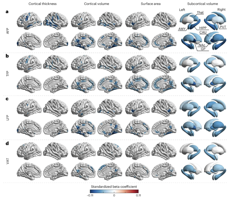
That’s obviously a lot for most of us to take in.
But the key message from the research is that visceral fat was the only type of fat directly linked to damage to the brain’s white matter – the wiring that carries signals between neurons.
White matter damage is closely tied to the biology behind vascular cognitive impairment and small vessel disease in the brain – conditions that sit upstream of dementia risk.
While the study didn’t look directly at what could be causing this link, the researchers think chronic, low-grade inflammation triggered by visceral fat may be spilling over into the brain itself, fuelling neuroinflammation.
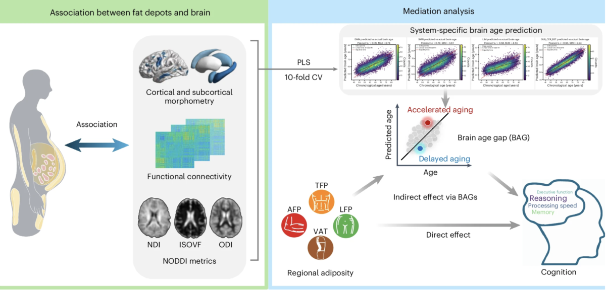
“Visceral adipose tissue secretes pro-inflammatory factors … triggering systemic inflammation that contributes to neuroinflammation and white-matter damage,” the team writes in their paper.
“In contrast, adiposity in the arms, trunk, and legs exhibits lower levels of inflammatory factor secretion.”
It’s a mechanism that lines up with the insulin and glucose pathway flagged by the team earlier this year.

The upside of this research is that how much body fat you have is something you can act on.
Of course, the research still has its limitations. While it looked at a large group, it’s still an observational study and hasn’t directly identified any causal factors.
Related: Whether You Carry Fat in Your Arms or Legs Could Predict Risk of Dementia
There’s still a lot more work to be done before we can fully unpack the relationship between this specific fat around our organs and our overall health. But the trend is increasing
“Regional adiposity is an intervenable and modifiable health risk factor,” says Qiu.
“Reducing visceral fat through targeted lifestyle interventions can open up new pathways for the proactive prevention and treatment of neurodegenerative diseases.”
The findings have been published in Nature Mental Health.
This article was fact-checked by Rebecca Dyer and edited by Rebecca Dyer. While we pride ourselves on our process, we are only human. If you spot a mistake, please let us know.