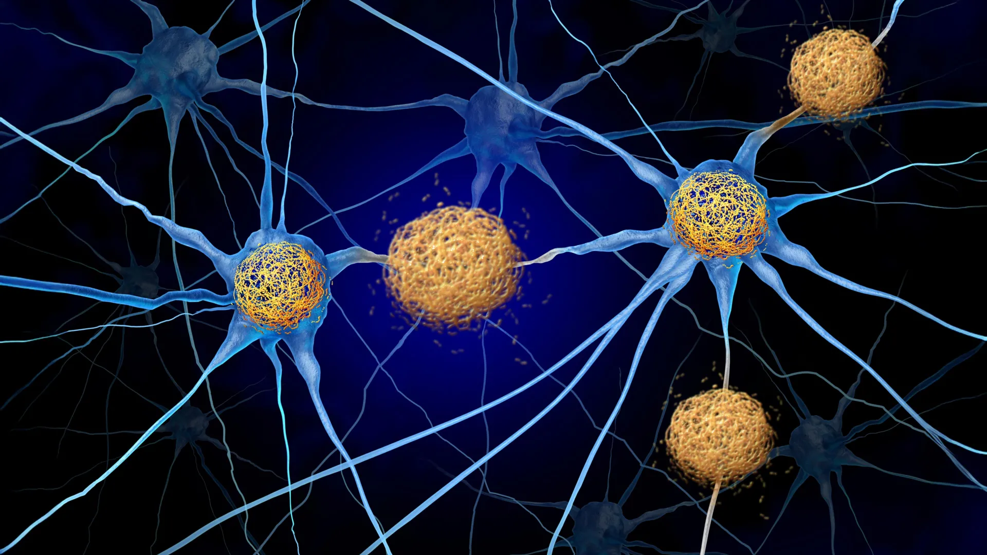
Тархины эд эсийг гэмтээдэг дархлааны хариу урвалыг саатуулснаар Альцгеймер болон түүнтэй төстэй өвчний явцыг удаашруулах боломжтойг судалгаагаар тогтоов.
Вашингтоны их сургуулийн Анагаах ухааны сургуулийн эрдэмтэд Альцгеймер болон бусад “тауопати” өвчний үед тархины эс үхэх үйл явцыг зогсоох шинэ стратегийг тодорхойлсон байна. Судлаачид “Neuron” сэтгүүлд нийтлэгдсэн судалгаандаа тархины эсэд хуримтлагдсан тау уураг өөрөө гол хор хөнөөл биш, харин түүнд хариу үйлдэл үзүүлж буй дархлааны тогтолцоо нь мэдрэлийн эсийг гэмтээхэд чухал үүрэгтэй болохыг тогтоожээ.
Туршилтын хүрээнд эрдэмтэд тау уургийн хуримтлал бүхий хулгануудад CXCR3 хэмээх уургийг хаах эсрэг биетийг тарьсан байна. Энэхүү уураг нь дархлааны T эсийг тархи руу нэвтрэхэд чиглүүлдэг гол хүчин зүйл юм. Эсрэг биет ашиглан T эсийн тархи руу орох урсгалыг хааснаар тархины эсүүд доторх дархлааны эсийн тоог хоёр дахин бууруулж чаджээ.
Туршилтын үр дүнд эмчилгээ хийлгэсэн хулгануудын ой санамжтай холбоотой тархины эд эс 40 орчим хувиар илүү хадгалагдаж, ой тогтоолтын сорил дээр илүү сайн үзүүлэлт үзүүлсэн байна. Сонирхолтой нь, энэхүү эерэг үр дүн нь тархин дахь хортой тау уургийн хэмжээнд ямар ч өөрчлөлт ороогүй байхад ажиглагдсан нь уг өвчнийг эмчлэх шинэ чиглэлийг нээж өглөө.
Судалгааны ахлах зохиогч, Анагаах ухааны доктор Дэвид М. Хольцман энэхүү арга нь тархины эсийг үхлээс хамгаалахад чухал ач холбогдолтой болохыг онцолжээ. Мөн эсрэг биет нь тархины эд рүү шууд нэвтрэх шаардлагагүй, зөвхөн тархины гаднах хил хязгаарт нөлөөлөхөд хангалттай байсан нь эмчилгээний шинэ боломжийг бий болгож байна. Энэ нь тархины цусан хангамжийн хаалтыг нэвтлэх шаардлагагүй тул ирээдүйд эм боловсруулахад томоохон давуу тал болох юм.
Хэдийгээр үр дүн нь ирээдүйтэй байгаа ч уг арга нь одоогоор зөвхөн хулгана дээр хийгдсэн тул хүн дээр аюулгүй, үр дүнтэй эсэхийг тогтоох нэмэлт судалгаа шаардлагатай байна. Гэсэн хэдий ч тау уургийг устгахыг оролдохын оронд дархлааны хортой хариу урвалыг хязгаарлах нь Альцгеймерийн өвчнийг эмчлэх шинэ стратеги болох төлөвтэй байна.
Дэлгэрэнгүйг эх сурвалжаас харах
↓Эх сурвалжийг нээх ↓
Protecting brain cells from destruction remains one of the biggest challenges in treating Alzheimer’s disease and related neurodegenerative conditions. These disorders, known as tauopathies, involve the buildup of abnormal tangles of a protein called tau inside the brain. As the tangles accumulate, nearby neurons begin to die. However, growing evidence suggests that tau itself may not be responsible for most of the damage. Instead, the immune system’s reaction to the protein appears to play a major role in the loss of brain cells, memory, and independence.
Scientists at Washington University School of Medicine in St. Louis have now identified a promising strategy for interrupting this damaging immune response. In mice with tau accumulation similar to that seen in Alzheimer’s, researchers used an antibody to block a protein called CXCR3, which helps immune cells known as T cells enter the brain. After several months of treatment, the number of T cells inside the brain had fallen by approximately half.
The results were striking. Compared with untreated mice, animals receiving the antibody retained roughly 40% more tissue in brain regions involved in memory and performed better on a memory test. These improvements occurred even though the amount of accumulated tau remained unchanged.
The findings were published September 28 in the journal Neuron.
“In tauopathies, including Alzheimer’s disease, there’s no treatment right now that actually decreases neurodegeneration,” said David M. Holtzman, MD, senior author of the study and the Barbara Burton and Reuben M. Morriss III Distinguished Professor in the Department of Neurology at WashU Medicine. “If we can show that we’re really decreasing brain cell death, it’s certainly worth trying to pursue that pathway to prevent the most devastating consequences of these diseases.”
Why Immune Cells May Be a Key to Alzheimer’s Brain Damage
Current Alzheimer’s treatments primarily focus on removing amyloid, another protein associated with the disease. Two approved medications, lecanemab and donanemab, target amyloid plaques that develop between brain cells during the early stages of Alzheimer’s. These deposits interfere with communication between neurons, contribute to the formation of tau tangles inside cells, and ultimately lead to neuronal death.
Although these medications can slow cognitive decline, they have not been shown to prevent brain cells from dying. They also offer no treatment for primary tauopathies, conditions in which tau accumulates without the presence of amyloid. Alzheimer’s is classified as a secondary tauopathy because both abnormal amyloid and tau are present.
Holtzman’s laboratory has been investigating whether the immune system offers another way to address these diseases. In a 2023 study published in Nature, the researchers discovered that T cells enter the brains of mice with abnormal tau accumulation, concentrating in regions containing the most tau and contributing to neurodegeneration.
A related study published earlier this month in Nature Neuroscience provided another important clue. It showed that the signals directing these T cells originate in lymph nodes located outside the brain.
“For this therapeutic approach, if it is safe, you wouldn’t have to design the drug to get into the brain — which is a big deal since most molecules don’t cross the blood-brain barrier well — and you don’t have to get rid of the tau to get this therapeutic effect.”
David Holtzman, MD, WashU Medicine
Scientists Identify the Pathway That Guides Immune Cells Into the Brain
Despite these earlier discoveries, researchers still needed to understand how activated T cells found their way into the brain.
T cells navigate through the body by following chemical signals known as chemokines. Earlier experiments in Holtzman’s laboratory had identified unusually high levels of one such signal, CXCL10, in mice with tau accumulation. Other researchers had also reported elevated CXCL10 levels in people with Alzheimer’s disease.
Activated T cells carry a surface receptor called CXCR3 that recognizes CXCL10, allowing the cells to follow its chemical signals. This relationship suggested that the CXCL10 and CXCR3 pathway might be responsible for guiding harmful T cells into the brain.
To investigate, the researchers studied mice that developed abnormal tau deposits resembling those associated with Alzheimer’s. The team included co-senior author Jason Ulrich, PhD, a professor of neurology at WashU Medicine, and first author Joshua T. Emmerson, PhD, a postdoctoral researcher in Holtzman’s laboratory.
The experiments revealed that T cells could not infiltrate the brain when mice lacked either CXCL10 or its matching receptor, CXCR3, on T cells. This protection persisted even when the scientists deliberately triggered inflammation to stimulate an immune response.
Experimental Treatment Preserves Brain Tissue and Memory
With the pathway identified, the team tested whether blocking CXCR3 with an antibody could reduce brain damage.
The researchers selected young mice that already had tau deposits in their brains but had not yet experienced substantial loss of brain cells. They administered the antibody once every five days for three and a half months.
By the end of the experiment, treated mice had approximately half as many T cells in their brains as untreated animals. They also showed less evidence of neuronal injury and retained substantially more brain tissue, including roughly 40% more tissue in memory-related regions. Their performance on a memory test was also better.
Importantly, these protective effects occurred without any detectable reduction in tau tangles. The treated and untreated mice had similar amounts of abnormal tau, suggesting that limiting the immune response could reduce neurodegeneration even when the underlying protein deposits remain.
Another finding could have major implications for future drug development. The antibody reached the boundary of the brain but did not enter the brain tissue itself.
This suggests that it may be possible to reduce neurodegeneration by targeting immune activity from outside the brain. Such an approach could avoid one of the major difficulties in developing neurological treatments: getting drugs across the blood-brain barrier, which prevents many substances from entering brain tissue.
A Potential New Direction for Alzheimer’s Treatment
Although the results are encouraging, the treatment has so far been tested only in mice. Additional research will be needed to determine whether the approach is safe and effective enough to evaluate in people.
Holtzman suggested that medications already used to control T cells in other diseases could provide a starting point. Certain treatments for multiple sclerosis and other autoimmune conditions, in which the immune system mistakenly attacks healthy tissue, might be worth investigating for their potential to reduce Alzheimer’s-related brain damage.
This would represent a significant shift in treatment strategy. Rather than concentrating exclusively on the abnormal proteins associated with neurodegeneration, researchers could also target the immune responses that contribute to the destruction of neurons.
“Tauopathies aren’t thought of as autoimmune disorders, so they haven’t been treated the same way, but this study shows for the first time in an animal model that these diseases respond to a specific T-cell therapy,” said Holtzman, who also directs WashU’s Hope Center for Neurological Disorders and the Knight Alzheimer Disease Research Center at WashU Medicine.
“For this therapeutic approach, if it is safe, you wouldn’t have to design the drug to get into the brain — which is a big deal since most molecules don’t cross the blood-brain barrier well — and you don’t have to get rid of the tau to get this therapeutic effect.”
The findings raise the possibility that protecting the brain from harmful immune activity, rather than removing tau itself, could become an important strategy for treating Alzheimer’s and other tauopathies.
The research received support from a BrightFocus Postdoctoral Fellowship, National Institutes of Health/National Institute on Aging grants R01AG082328 and R01AG085374, the GHR Foundation, the Carol and Gene Ludwig Initiative in Neuroimmunology Research, a gift from Ronald Schaich, a gift from John Ludwig, a gift from Cindy and Evan Goldberg, and the Freedom Together Foundation. The study’s content is the responsibility of its authors and does not necessarily reflect the official views of the NIH.
Disclosures: DMH co-founded C2N Diagnostics and serves on its scientific advisory board. DMH also serves on the scientific advisory boards of Denali, Genentech, and Switch; provides consulting services to Pfizer, Roche, Novartis, Annexon, and Acta; and serves on the advisory boards of Neuron and Cell.