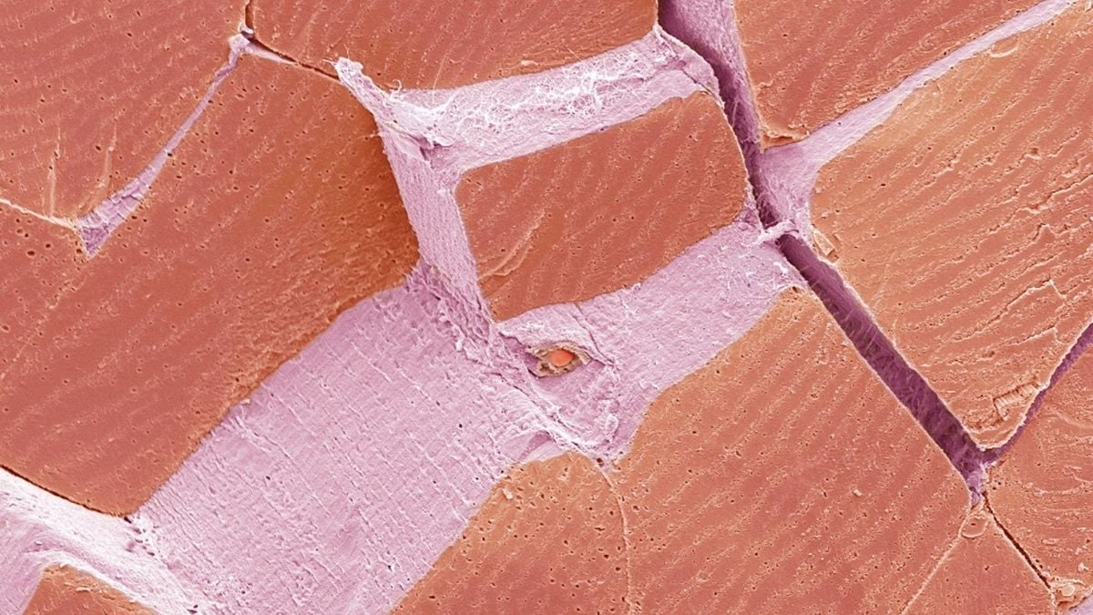
Шинжлэх ухааны судалгаанд ашигладаг электрон микроскопоор авсан энэхүү зурагт ягаан болон улаан өнгөөр ялгарч буй хэсгүүд нь булчингийн ширхэгүүд юм. Эдгээр ширхэг нь нэгдэж булчингийн багцыг үүсгэдэг бөгөөд тэдгээрийн хоорондох нил ягаан өнгийн торлог бүтэц нь булчингийн эд эсийг холбож, цусны судас, мэдрэлийн төгсгөлүүдийг агуулдаг перимизиум (perimysium) хэмээх холбогч эд ажээ.
Зурган дээрх жижиг зууван хэлбэртэй бүтэц нь хялгасан судас бөгөөд энэ нь булчингийн үйл ажиллагаанд шаардлагатай хүчилтөрөгчийг тасралтгүй дамжуулдаг. Электрон микроскопоор авсан анхны дүрс нь хар цагаан өнгөтэй байдаг ч судлаачид булчингийн бүтэц, цусны судас болон эд эсийг ялгахад хялбар болгох үүднээс дараа нь өнгө нэмдэг байна.
Энэхүү судалгаа нь хүний булчингийн нөхөн төлжилтөд чухал үүрэгтэй “хиймэл дагуул” эсүүдийн ач холбогдлыг тодотгож байна. Эдгээр эс нь хөдөлгөөний идэвхтэй байдлаас хамааран булчингийн эдийг нөхөн сэргээдэг боловч удаан хугацаанд идэвхгүй байвал хуваагдах чадвараа алддаг болохыг эрдэмтэд онцоллоо.
Дэлгэрэнгүйг эх сурвалжаас харах
↓Эх сурвалжийг нээх ↓
If you’re a reader of our weekly newsletter, you’ll know that one of our most popular features is our Microscope Mystery.
Each week, our team shares a head-scratching microscopic image and asks readers to work out what exactly they’re looking at. Occasionally with hilarious results.
(Like the time our assistant editor Clare Watson guessed that the surface of a solar panel was actually a biscuit – “or maybe I’m just hungry” – or journalist Jess Cockerill said the scent glands of a rose petal looked like “my tastebuds after I burnt them on my morning coffee“.)
Due to popular demand, we will now be hosting these microscopic conundrums on the site, too, a few days after the newsletter.
Introducing: Monday Microscope Mystery!
Read on for this week’s puzzle, and don’t forget to compare your answers to those on socials.
To kick us off, we have this strangely tasty-looking pink and purple landscape.
What do you think you’re looking at?
We’ll put in some crazy footage of orcas punching sunfish so hard that they explode below as a visual buffer.
There will be spoilers below, you have been warned!

Narrow it down…
Some ideas to narrow down what the image could be of:
a. Ocean sponge
b. Shark scales
c. Skeletal muscle
d. Lizard tongue
We’ll show you the image one more time and will reveal the answer below.
Okay ready?
You sure?
Alright… here we go.
The answer
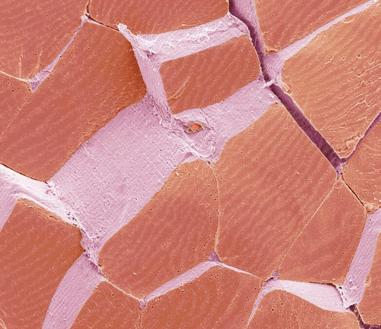
The answer is C: skeletal muscle.
Each of those blocky reddish-pink shapes is a single muscle fiber – a cell so long and specialized it can stretch the entire length of some muscles in your body.
Bundle a few hundred of these fibers together and you get a fascicle.
Bundle a few hundred fascicles together and you get a muscle you’d actually recognize, like a bicep or a hamstring.
The light purple webbing running between the fibers is connective tissue called perimysium, and it’s doing more than just holding cells together. It’s shot through with blood vessels and nerve endings, wrapping each fiber so the whole bundle can contract in sync without shearing itself apart.
Look closely at the pink band, just above center, and there’s a tiny textured oval peaking out.
That’s a capillary.
It’s one of the thousands of microscopic vessels threading through muscle tissue, delivering the oxygen that fibers burn through the moment you start moving.

None of this colour is real, by the way.
This image was captured with a scanning electron microscope, which fires a beam of electrons at the sample instead of light, producing a black-and-white image far more detailed than any optical microscope could manage. The reds and pinks are added afterward, partly for beauty, partly to help the eye tell muscle from tissue from vessel at a glance.
Related: Good News, We Can Activate The Cells That Keep Our Muscles From Wasting Away After 30
The magnification is 3,000x when printed at 10 centimeters (3.9 inches) wide.
What’s wild is that skeletal muscle relies on satellite cells to regenerate. But these cells only stay primed for the job if you keep moving. Left dormant too long, they can lose the ability to divide at all.
So next time you flex or find yourself shivering to stay warm, you’re looking at a tiled, wired, self-repairing machine built from thousands of fibers working in perfect unison – just one intense zoom away from looking exactly like a cracked-tile floor.
And don’t forget to sign up to our newsletter for more Microscope Mysteries!
This article was fact-checked by Clare Watson and edited by Clare Watson. While we pride ourselves on our process, we are only human. If you spot a mistake, please let us know.