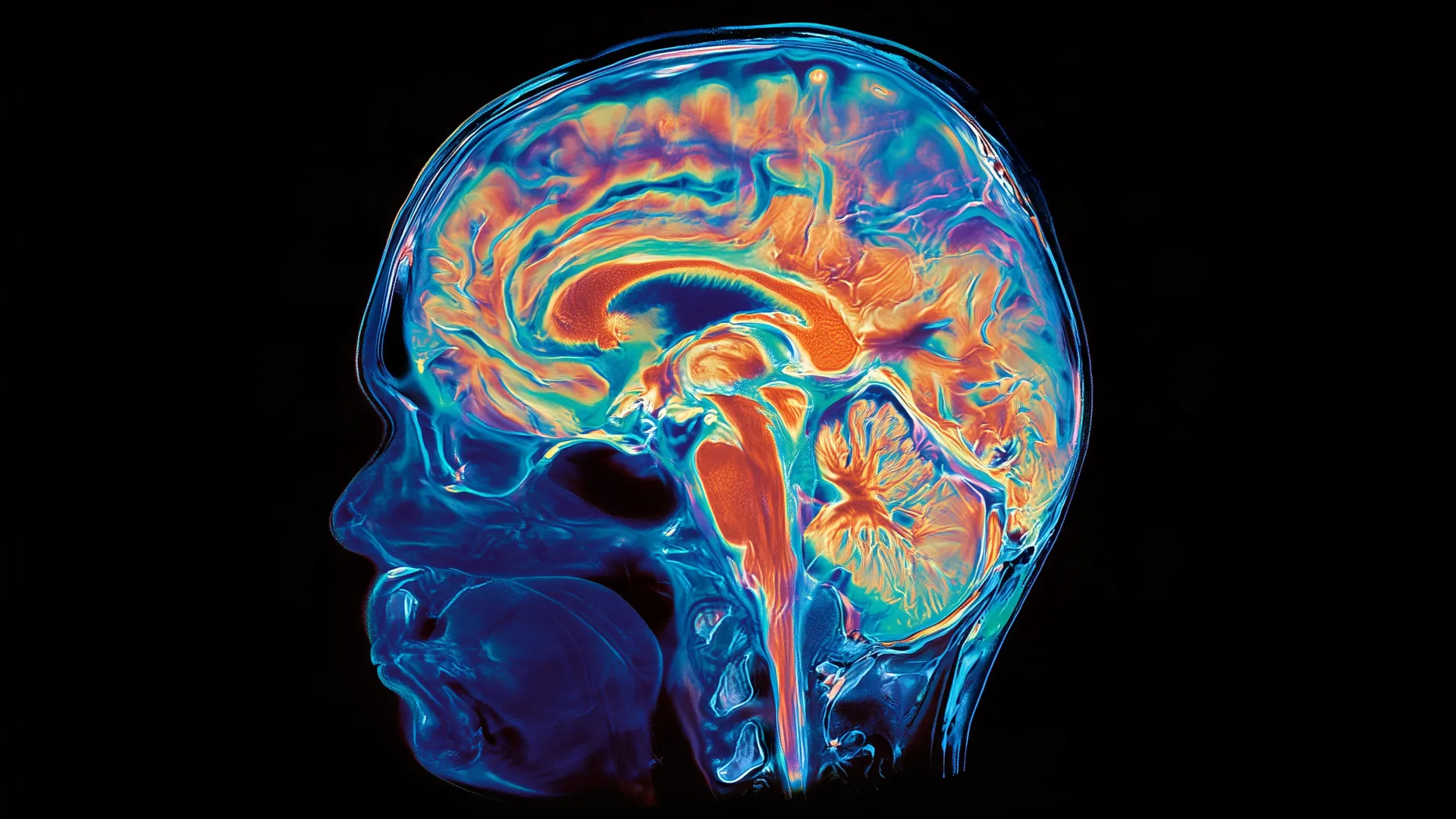
Судлаачид хүний тархины хөгжил нь нэг эх үүсвэрээс бус, харин хувьслын явцад тусдаа хөгжсөн хоёр өөр систем нэгдсэнээр үүссэн болохыг илрүүлжээ.
Стэнфорд Медициний эрдэмтдийн “Nature Neuroscience” сэтгүүлд нийтлүүлсэн судалгаагаар хүний тархи нь урьд өмнө төсөөлж байснаас ялгаатай бүтэцтэй болохыг тогтоов. Уламжлалт онолоор тархийг нэг эх үүсвэрээс хөгждөг нэгдмэл эрхтэн гэж үздэг байсан бол шинэ судалгаагаар тархины урд хэсэг болон хойд хэсэг (тархины үүдэл) нь хөгжлийн эхний үе шатнаасаа эхлэн тус тусдаа эсийн бүлгээс үүсдэгийг нотолжээ.
Судлаачид хулганы үр хөврөл дээр хийсэн туршилтаар тархины урд хэсгийг бүрдүүлэгч “Otx2” ген болон хойд хэсгийг бүрдүүлэгч “Gbx2” генийн илрэл бүхий эсийн популяцууд огтлолцолгүй, тусдаа замаар хөгждөгийг илрүүлсэн байна. ДНХ-ийн савлагаа буюу хроматины бүтэц нь ч энэ хоёр бүсэд үндсээрээ ялгаатай байгаа нь эдгээр эсүүд хөгжлийн явцад эртнээс тусгаарлагдсаныг гэрчилж байна. Энэхүү ололт нь өмнө нь эрдэмтдийн хувьд хүндрэлтэй байсан тархины үүдлийн мэдрэлийн эсийг лабораторийн нөхцөлд гарган авах боломжийг нээж өгчээ.
Хувьслын түүхийг судлахад 550 гаруй сая жилийн өмнө амьдарч байсан амьтдад ч тархины ижил хоёр эх үүсвэртэй зохион байгуулалт ажиглагддаг нь тогтоогдсон. Энэхүү нээлт нь нугасны булчингийн хатингаршил (SMA) болон хажуугийн хатингаршилт склероз (ALS) зэрэг тархины үүдэлтэй холбоотой ноцтой өвчнийг судлах, нөхөн сэргээх эмчилгээг хөгжүүлэх шинэ арга замыг тодорхойлж байна. Одоо судлаачид нугасны хөгжлийн эх үүсвэр болон дээрх өвчнүүд тархины үүдлийн эсүүдэд хэрхэн нөлөөлдгийг нарийвчлан судлахаар төлөвлөж байна.
Дэлгэрэнгүйг эх сурвалжаас харах
↓Эх сурвалжийг нээх ↓
Scientists have long treated the brain as a single organ with a shared developmental origin. New research led by Stanford Medicine challenges that basic assumption, suggesting that what we call the brain is actually built from two distinct systems that arose separately over hundreds of millions of years of evolution.
For decades, the dominant model held that the entire brain develops from one early progenitor cell population. Under that view, the forebrain, midbrain, and hindbrain all trace back to the same developmental starting point.
The new findings point to a very different picture. Researchers found that the human brain appears to combine two ancient nervous systems. One gives rise to structures responsible for essential functions such as breathing and controlling the heartbeat. The other produces regions associated with abilities such as language, mathematics, abstract thought, and reflection on our own existence.
The discovery may also explain a long-standing problem in neuroscience: why researchers have had so much difficulty growing certain brain cells in the laboratory. It could create new opportunities to investigate diseases involving the brain stem, including spinal muscular atrophy (also known as SMA) and amyotrophic lateral sclerosis (also known as ALS or Lou Gehrig’s disease).
“We’ve shown for the first time that the front of the brain arises from a totally different progenitor cell than the back of the brain,” said Kyle Loh, PhD, associate professor of developmental biology. “Our discovery means that we can now grow neurons from the back of the brain, the hindbrain, in a petri dish and study their functions.”
The findings will be published in Nature Neuroscience Sept. 18. Loh is the senior author. Graduate students Carolyn Dundes and Rayyan Jokhai are co-first authors of the research.
Two Developmental Origins of the Brain
The adult brain is divided into three major regions: the forebrain, midbrain and hindbrain. The forebrain supports many of the abilities people most closely associate with complex thought, including language, consciousness and abstract reasoning.
The hindbrain, by contrast, sits toward the back of the skull and is often referred to as the brain stem. It helps control automatic processes essential for survival, including breathing, sleeping, heartbeat regulation and hunger. Hindbrain neurons also direct muscles in the face, tongue and throat that are important for speaking and swallowing.
Despite the hindbrain’s vital role, scientists have spent decades struggling to create human hindbrain neurons in the laboratory. That limitation has made it much harder to study serious diseases involving the brain stem, including spinal muscular atrophy and amyotrophic lateral sclerosis.
SMA is a leading genetic cause of death in children under 1 year of age. ALS is often diagnosed between the ages of 40 and 70 and affects both the forebrain and the hindbrain. In both diseases, particular hindbrain neurons progressively stop functioning. Patients can eventually lose the ability to swallow, increasing the risk of pneumonia if food or liquid enters the lungs, and later may lose the ability to breathe.
A Split That Begins Early in Development
The key insight came when the researchers examined one of the earliest stages of embryonic development. During gastrulation, the basic structure of the body begins taking shape.
Jokhai and Dundes found that the hindbrain does not emerge as a later branch of the developmental pathway that produces the forebrain and midbrain. Instead, it follows its own pathway from the beginning, developing in parallel with the other regions.
To investigate this process, the researchers studied developing mouse embryos and identified two different populations of brain progenitor cells.
One population expresses a gene called Otx2 and is destined to form the forebrain and midbrain. A second population expresses a gene called Gbx2 and develops into the hindbrain. The researchers found that the two populations remain separate and do not overlap, even at the earliest stages they examined.
That distinction became even clearer when the team studied chromatin, the material that packages DNA inside cells and helps control which genes are available for use and which remain inaccessible.
The anterior neural ectoderm (future forebrain and midbrain) and posterior neural ectoderm (future hindbrain) had fundamentally different chromatin configurations. Those differences effectively committed the cells to separate developmental paths from the start.
“Previous attempts to make hindbrain neurons likely tried to coax forebrain and midbrain progenitors into hindbrain cells, which our study shows is not possible,” Jokhai said.
The finding may explain why earlier attempts to make hindbrain neurons proved so difficult. Researchers may have been trying to convert one type of progenitor cell into another fate that it was never capable of adopting.
“In stem cell biology, people are always fixated with creating the end cell type, like the neuron,” Jokhai said. “But it’s important to begin at the earliest stages of embryonic development. Our careful attention to that early time point allowed us to find this fundamental split in brain development.”
Growing Human Hindbrain Neurons in the Lab
Once the researchers understood that the hindbrain starts from a separate developmental pathway, they were able to use that knowledge to solve a major laboratory challenge.
For the first time, the team successfully guided human pluripotent stem cells (a kind of cell that can create any cell in the human body) into becoming functional hindbrain motor neurons.
The cells behaved like genuine hindbrain neurons. They produced electrical signals known as action potentials and made proteins associated with specific regions of the hindbrain that control facial and swallowing muscles.
The researchers then turned to evolution to see how far back this developmental split might extend.
A Pattern More Than 500 Million Years Old
Looking across more than 550 million years of evolutionary history, the team found evidence of the same two-origin arrangement in chickens, zebrafish and even acorn worms. These small animals live on the seafloor and share a very distant common ancestor with humans.
The researchers also noted that jellyfish, which diverged from humans about 600 to 700 million years ago, possess two nervous systems positioned at different ends of their bodies.
Together, those findings raise the possibility that the modern vertebrate brain arose when evolution brought two preexisting neural systems into close physical proximity.
“Our research suggests that evolution took two existing neural systems and pushed them together spatially,” Loh said. “Having the brain as one organ would probably be more efficient, but we rely on this primordial way to make the brain as two separate pieces.”
“I was surprised at our findings because the word ‘brain’ implies a contiguous organ that likely has a singular origin,” Jokhai said. “But even 500 million years ago, there were these separate neural systems, which now almost operate as one, which is very cool.”
New Possibilities for ALS and SMA Research
The work could have important implications for scientists studying SMA, ALS and other disorders that damage the brain stem.
One major obstacle has been access to human tissue. Brain stem samples cannot be collected from living patients for routine research, making it extremely difficult to examine affected neurons directly. The ability to grow hindbrain neurons in a dish could allow scientists to study what goes wrong in these diseases with far greater precision.
The hindbrain is also involved in another major area of medicine: weight control. It contains neural circuits that regulate hunger, which are among the systems influenced by weight-loss drugs such as semaglutide.
The researchers now want to investigate where the spinal cord comes from developmentally and determine more precisely how SMA and ALS disrupt hindbrain neurons.
“Now we have a model to better understand these devastating diseases, and work toward regenerative therapies for them,” Jokhai said. “This is a very exciting new frontier in brain research.”
Researchers from the California Institute of Technology and the University of California, San Francisco contributed to the study.
This work was supported by the National Institutes of Health (grants DP5OD024558, DP2GM146258, R00GM121852, R01DK115728, R01DE027538, T32GM119995, T32GM007365, T32GM007790 and F31DE031154); the National Science Foundation; the California Institute for Regenerative Medicine; the Spinal Muscular Atrophy Foundation; a Stanford Maternal and Child Health Research Institute grant; the Stanford Beckman and Ludwig Centers; the Siebel Stem Cell Institute; a Stinehart-Reed Foundation grant; the Gatsby Charitable Foundation; the Howard Hughes Medical Institute; the Packard Foundation; the Pew Charitable Trusts; the Baxter Foundation; the Human Frontier Science Program; and the anonymous, Fickel, Gilbert, and Stinehart-Reed families.