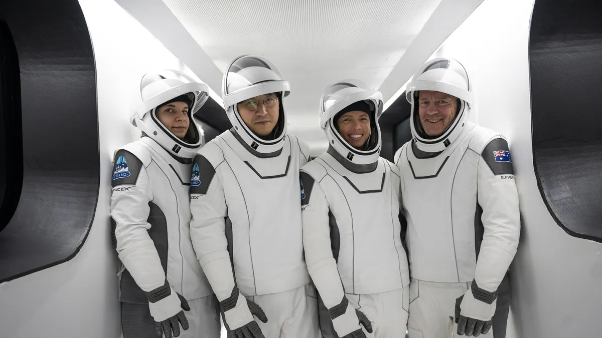
Олон улсын судлаачдын баг арилжааны сансрын нислэгээр дамжуулан сансарт анх удаа рентген шинжилгээг амжилттай гүйцэтгэж, сансрын анагаах ухаанд томоохон дэвшил авчирлаа.
Сансрын анагаах ухаанд сүүлийн дөчин жилийн турш зөвхөн хэт авианы төхөөрөмжийг найдвартай дүрс оношилгооны хэрэгсэл болгон ашиглаж ирсэн. Гэвч урт хугацааны болон алсын зайн сансрын аялал нэмэгдэхийн хэрээр илүү өргөн хүрээний эрүүл мэндийн асуудлыг шийдвэрлэх чадвартай, найдвартай оношилгооны хэрэгцээ улам бүр өсөж байна. Майо Клиникийн сансрын анагаах ухааны профессор Шейна Гиффордоор ахлуулсан олон улсын эрдэмтдийн баг энэхүү хязгаарлалтыг даван туулах зорилгоор зөөврийн рентген системийг сансарт туршжээ.
Тус багийнхан SpaceX-ийн Fram2 арилжааны сансрын нислэгийн үеэр хөнгөн жинтэй, утасгүй дижитал рентген үүсгүүр бүхий системийг ашиглан хүний биеийн болон тоног төхөөрөмжийн дүрслэлийг авсан байна. Сансарт хөөрөхийн өмнө багийн гишүүд зөөврийн рентген системийг ашиглах талаар дөрвөн цагийн сургалтад хамрагдсан бөгөөд нислэгийн үеэр гар, шуу, хэвлий, аарцаг болон цээжний рентген зураг авчээ. Бие даасан радиологичдын үнэлснээр, сансарт авсан эдгээр зургийн чанар, дүрслэл нь оношилгоонд ашиглахад бүрэн тохиромжтой болох нь тогтоогдсон байна.
Энэхүү судалгаа нь сансрын хөлөг дээр рентген оношилгоо хийх боломжтойг баталснаар сансрын нисгэгчдийн эрүүл мэндийг хамгаалахаас гадна сансрын хөлгийн эд анги, электроник болон сансрын хувцасны гэмтлийг задлахгүйгээр шалгах чухал ач холбогдолтой юм. Хэдийгээр цээж, аарцагны байрлал тогтоох үед бага зэргийн зөрүү гарсан ч ерөнхий нарийвчлал нь оношилгооны стандартад нийцжээ. Цаашид энэхүү зөөврийн системийг улам боловсронгуй болгож, жижиг хэмжээтэй, илүү бат бөх болгосноор ирээдүйн сансрын нислэгүүд болон сарны гадаргууг шинжлэх судалгаанд өргөнөөр ашиглах боломж нээгдэж байна.
Дэлгэрэнгүйг эх сурвалжаас харах
Эх сурвалжийг нээх ↓
With many plans for crewed missions to the Moon and Mars, and commercial space flights becoming more common each year, there is a growing need to develop medical procedures that will address the health hazards of space. Aside from the usual concerns regarding radiation exposure, bone density, and muscle loss, diagnostic tools must also be available to determine the extent of injuries and ailments. Developing these and ensuring astronaut health and longevity are the objectives of the growing field of “space medicine.”
A major milestone was recently achieved when a team of international, interdisciplinary researchers conducted the first X-ray diagnostic during a commercial spaceflight. This is a major step in space medicine because ultrasound has been the only reliable medical imaging tool for the past four decades, but it has limitations. As spaceflight missions increase in duration and distance, the need for reliable diagnostic tools that can address a wide range of health problems will only grow.
The research was led by Dr. Sheyna Gifford, a professor of aerospace medicine at the Mayo Clinic. It included radiologists, technicians, and engineers from the University of California San Diego (UCSD), Stanford University, Radiology Logistics Consultants Seattle, the Massachusetts Institute of Technology’s (MIT) Institute for Medical Engineering and Science, MinXray, KA Imaging, the University of Waterloo, Space Exploration Technologies, and more. The results of their mission appeared in the journal Radiology.
JAXA (Japan Aerospace Exploration Agency) astronaut Satoshi Furukawa pedals on the upgraded CEVIS system, keeping muscle and bone density levels during spaceflight. (Credit: NASA)
For logistical reasons, space medicine and whole-body medical imaging have been limited to ultrasound for more than four decades. Whereas ultrasound can be performed with handheld devices and imaging equipment, X-ray imaging traditionally required large machines that would increase the radiation dose astronauts receive. As Gifford explained in a recent press release by the Radiological Society of North America (RSNA):
It’s been a dream for aerospace medicine to have more than one imaging modality for diagnosing illnesses and injuries in space. X-rays are fast, easy, and diagnostically valuable. Traditional X-ray machines are very large, produce a lot of radiation, and have a tendency to produce a blurred image if there’s movement. Because everything in space is constantly moving, the conceit has been that obtaining a diagnostic image in orbit was too technically challenging.
The situation has changed in recent years with the creation of commercial off-the-shelf, portable X-ray machines, which are drastically increasing access to this medical technology. Beyond portable medicine in underserved communities and developing nations, space medicine is another potential application. In 2022, Gifford and her team investigated the feasibility of using a portable radiography system during a parabolic flight that simulated the microgravity of spaceflight. As she said:
Portable X-ray machines are in use everywhere—at the Kentucky Derby, on the sidelines of the Super Bowl and around the globe in low-resource areas—because they can run on solar power and can be operated by individuals with no medical expertise. We believed an off-the-shelf portable system would stand a very good chance of surviving prelaunch testing and be operational in space by crew members with minimal training.
Before the flight, three crew members spent four hours training on how to use the portable radiography system while SpaceX personnel conducted impact and compatibility testing on the system for the spacecraft. Despite the challenges, the crew successfully obtained a digital X-ray of a hand in simulated microgravity. Building on this, Dr. Gifford and her team partnered with SpaceX to test their portable X-ray system during Fram2, a commercial spaceflight mission sponsored by entrepreneur Chun Wang.
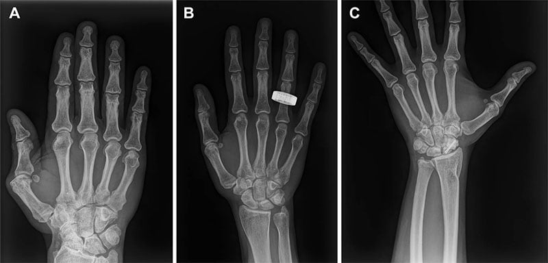 X-ray images obtained by Dr. Gifford and her team during the Fram2 commercial spaceflight. Credit: Gifford, S.A. et al. (2026)
The mission was a first to send a spacecraft into polar orbit, where it remained for just over three and a half days. Said Gifford:
A spaceflight-ready radiography system would have profound implications not only for crew health but also for mission-critical nonmedical tasks. For sustained human presence in space, X-rays are critical not just for crew members but also for other mission components like electronics and spacesuits. The only way to look inside these objects without taking them apart is to X-ray them.
In the study, the team used an X-ray system featuring an ultraportable wireless digital X-ray generator to obtain anatomic and equipment scans before and during flight. This included X-ray images of a hand, forearm, abdomen, pelvis, and chest. After acquiring X-rays of a phantom object to calibrate the system, the crew acquired in-flight images of the same anatomical parts. The crew also acquired postflight X-rays replicating the preflight and in-flight images upon return.
All of the X-ray images were evaluated by three independent radiologists, who found no differences in the overall image quality. Although they received lower scores for chest, pelvis, and abdomen positioning, the overall spatial resolution, contrast resolution, and positioning were consistent with diagnostic X-rays. The crewmembers also stated that the X-ray system was easy to use and the protocol easy to follow. As Gifford summarized:
By acquiring the first human and equipment X-rays in space, our study demonstrates the feasibility of in-orbit radiography and expanded diagnostic capabilities for crew health and hardware evaluation. Acquiring diagnostically useful X-rays in space is something that anyone can do. Three very talented nonmedical people with four hours of training in one of the harshest environments did it right and did it well.
Beyond space medicine, the development of portable X-ray systems extends to other space-related applications, including the imaging of malfunctioning satellites in orbit and equipping lunar rovers to analyze the Moon’s surface.
“It’s my hope that we can further reduce the size of portable imaging systems and improve its ruggedness and usability so they can be included in future missions,” added Gifford. “Disseminating autonomous miniature X-ray systems around the globe could also change the game in public health,” she added. “The sky is not the limit when it comes to X-rays in space and here on Earth.”