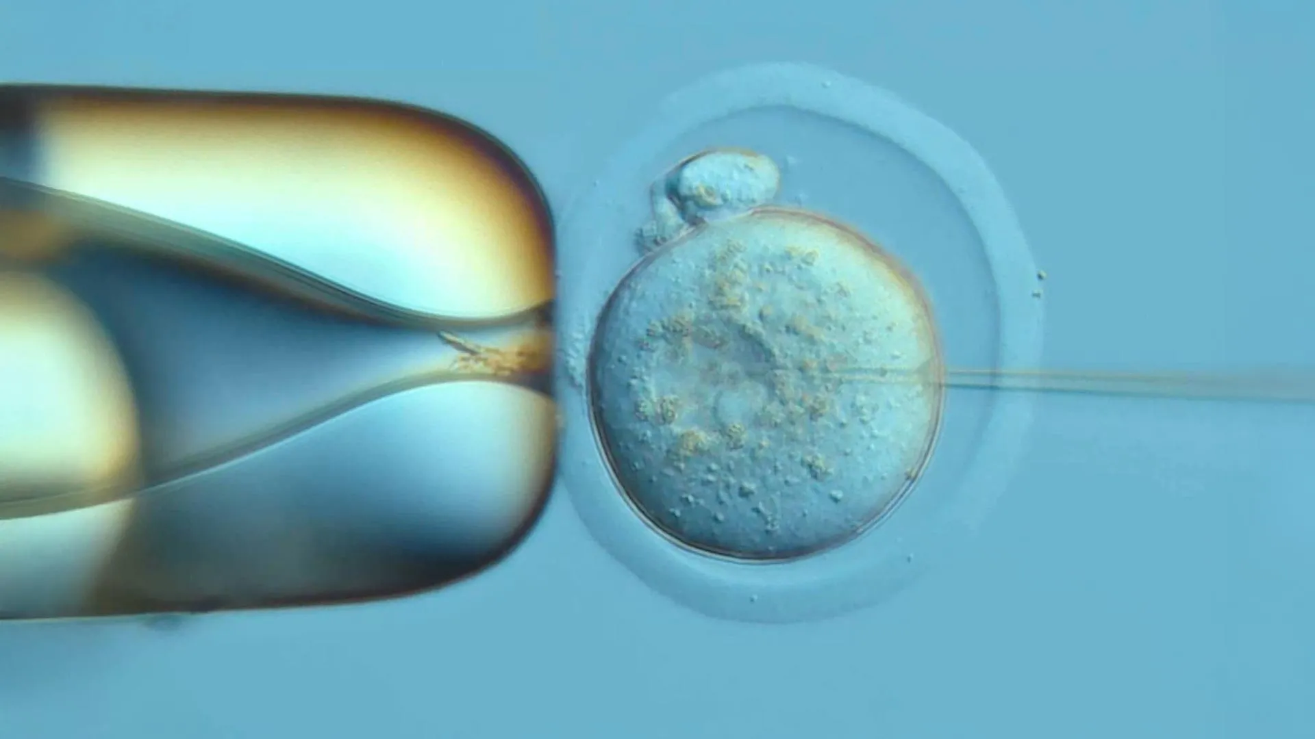
Эрдэмтэд хулганы үр хөврөлийн анхны эсээс бие махбод бүрэлдэх хүртэлх үйл явцыг ДНХ-д мэдээлэл хадгалах шинэлэг аргаар хянаж, эсийн удмын санг нарийвчлан тогтоожээ.
Вашингтоны их сургуулийн Анагаах ухааны сургуулийн судлаачид “DNA Typewriter” хэмээх технологийн тусламжтайгаар үр тогтсон өндгөн эс анх хоёр хуваагдсанаас хойшхи 13.5 хоногийн турш эсүүдийн хөгжлийн түүхийг мөшгөн судалсан байна. “Science” сэтгүүлд 2026 оны аравдугаар сарын 8-нд нийтлэгдсэн энэхүү судалгаа нь эсүүд хэрхэн үржиж, тусгай үүрэг гүйцэтгэх болсныг биологийн тэмдэглэлийн дэвтэр мэтээр ДНХ-д нь хадгалах боломжийг олгожээ. Судлаачид хулганы үр хөврөлийн геномд “соронзон хальс” мэт үүрэг гүйцэтгэх ДНХ-ийн дарааллыг суулгаснаар эс хуваагдах бүрт шинэ мэдээлэл бичигдэж, улмаар эсийн удмын холбоог нөхөн сэргээх боломжтой болсон байна.
Судалгааны явцад үр тогтсон өндгөн эсийн анхны хоёр эс нь тус бүртээ биеийн олон төрлийн эсийг бүрдүүлэхэд ижил хувь нэмэр оруулдгийг илрүүлжээ. Эсийн хөгжлийн явцыг ажиглахад цусны эсүүд болон нүдний торлог бүрхэвч харьцангуй эрт үүрэг чиглэлээ олдог бол арьсны эсүүд хожим нь мэргэшдэг болох нь тогтоогдсон юм. Энэхүү үйл явц нь эсийн хөгжил нэгэн зэрэг явагддаггүй, харин эд эрхтэн бүр өөрийн гэсэн цаг хугацааны хуваарьтай болохыг харуулж байна.
Өмнө нь хэрэглэж байсан эсийн хөгжлийг судлах аргууд нь ДНХ-ийг таслах замаар тэмдэглэгээ хийдэг байсан нь эсийг гэмтээх эсвэл өмнөх мэдээллийг устгах сул талтай байв. Харин “DNA Typewriter” нь ДНХ-ийг бүрэн таслахгүйгээр дарааллаар нь бичлэг хийдэг тул илүү өндөр нарийвчлалтай мэдээлэл өгдөг аж. Энэхүү ололт нь төрөлхийн гажиг, мэдрэлийн хөгжлийн эмгэг, хорт хавдар болон үүдэл эсийн эмчилгээний судалгаанд чухал ач холбогдолтой юм.
Дэлгэрэнгүйг эх сурвалжаас харах
↓Эх сурвалжийг нээх ↓
Scientists have reconstructed a remarkably detailed family tree of cells in a developing mouse embryo, tracing their origins back to the moment a fertilized egg first divided into two cells. Using an innovative DNA recording technology, the researchers followed how those early cells multiplied, developed specialized roles, and eventually helped form a body with distinct organs and tissues.
The recording covered the first 13.5 days of embryonic development, providing an unusually detailed look at how a complex mammalian body takes shape. (Full gestation in mice is 19 to 21 days.)
The findings, published on October 8, 2026, in the journal Science, could help scientists better understand normal development and what happens when that process goes wrong.
“We are interested in tracking how cells’ early ancestry in an embryo influences their later fates. Exploring cell lineages helps decode normal development. Such research could eventually advance knowledge about congenital malformations, neurodevelopmental conditions, genetic disorders or cancer,” said Dr. Jay Shendure, a professor of genome sciences at the University of Washington School of Medicine in Seattle and a Howard Hughes Medical Institute Investigator.
Shendure led the research alongside Dr. Chengxiang Qiu, a molecular and systems biologist at Dartmouth College in New Hampshire. Both served as senior authors of the study.
A DNA Typewriter Records the History of Living Cells
The achievement was made possible by a technology called DNA Typewriter, developed by Shendure and Junhong Choi, who is now at Sloan Kettering Cancer Center.
Rather than simply observing cells as they divide, the system allows scientists to preserve a record of their history directly within their DNA.
To accomplish this, the researchers inserted a redesigned recording sequence, which they call a tape, into the genome of a fertilized mouse egg. The updated design made it easier to recover the information stored in individual cells.
As the embryo developed, the DNA Typewriter remained embedded in the genetic material of its cells, recording their divisions in a way that could later be used to reconstruct their ancestry. The result was something like a biological logbook, documenting the sequence of events that gave rise to different cell populations.
“Think of it like an actual typewriter, except it types onto a cell’s own DNA instead of paper,” explained Haedong Kim, a co-first author of the study and a postdoctoral scientist in genome sciences at UW Medicine, the Seattle Hub for Synthetic Biology and the Howard Hughes Medical Institute. “Every time a cell divides, it strikes one new character onto the next blank line — always in order, never overwriting what’s already there. Because the characters are always filled in sequence, that sequence itself spells out the cell’s division history.”
Because these markings are passed down to daughter cells, researchers can examine the recorded sequences and identify which cells descended from a shared ancestor. Cells carrying matching portions of the record can be linked together, allowing scientists to reconstruct branches of the embryo’s family tree.
“Writing the record is only half the problem. You also have to read it back out of each cell, so we redesigned the tape to make that easier,” said Kim.
Overcoming a Major Challenge in Developmental Biology
Reconstructing the entire history of a developing mammal has long been a formidable challenge. As an embryo grows, its cells divide repeatedly and begin taking on specialized functions. Following those relationships becomes increasingly difficult as the number and variety of cells expand.
Scientists accomplished a similar feat decades ago in a tiny roundworm, but that animal has a relatively simple body with far fewer cells. Its transparency also makes development easier to study.
Mammals are considerably more complicated. Earlier technologies provided important insights into mouse embryonic development, but they generally captured only portions of the larger cellular family tree.
Some traditional recording techniques also have significant limitations. They work by cutting DNA to create identifying marks, which can damage cells and erase information left by earlier divisions. Their recording capacity is limited, and because the marks are not necessarily created in a fixed sequence, scientists must attempt to reconstruct the order of events afterward.
“DNA Typewriter avoids all of this,” said Kim. “It writes without fully severing the DNA, keeps recording relatively steadily, and writes everything in strict order, so we can record cell lineages at much higher resolution for a longer time.”
Tracing an Embryo Back to Its First Two Cells
The researchers began by attempting to record development in 100 fertilized mouse eggs. Of those, 10 embryos were ultimately available for examination.
One specimen, identified as Embryo No. 3, stood out because its DNA recording system had functioned particularly well and preserved the most extensive developmental history. The scientists therefore concentrated their detailed analysis on that embryo.
One of the most important discoveries came from a recording made at the very beginning of development, immediately after the fertilized egg divided for the first time.
“Our recording captured a clear, permanent mark distinguishing the two very first cells, letting us trace nearly every cell we profiled from the embryo back to one or the other,” said Kim. “One of these two founding cells did go on to produce more descendants than the other, but they produced diverse cell types at almost equal ratios.”
This meant that although one of the original cells contributed more descendants overall, both founding cells gave rise to a remarkably similar mix of specialized cell types.
The finding illustrates how scientists can now investigate the earliest stages of development with a level of detail that was previously difficult to achieve.
Scientists Discover When Cells Choose Their Roles
The DNA recordings also revealed important information about when cells begin committing to their eventual roles in the body.
Early embryonic cells can give rise to many different types of tissue. As development progresses, their descendants gradually become more specialized, eventually forming structures such as the nervous system, skin, blood, and eyes.
By reconstructing the branches of the embryo’s cellular family tree, the researchers could determine when different groups of cells began following distinct developmental paths.
Their observations were consistent with conclusions drawn from decades of previous research, but the new approach allowed them to examine these patterns within a single developing mouse.
“Blood cells and the retina committed relatively early, while the skin’s outer layer didn’t commit until noticeably later,” the researchers explained. “Cell fate wasn’t locked in all at once. Different cell types settled into their identity on their own separate schedules, some early and some late.”
These findings show that the transition from a flexible, unspecialized cell to one with a particular biological function does not happen at the same time throughout the embryo.
Instead, different tissues follow their own developmental timelines. Some cells begin narrowing their possible futures early, while others remain flexible for a longer period before becoming committed to a specific role.
What This Could Mean for Birth Defects, Cancer, and Stem Cell Therapies
Understanding exactly how cells acquire their identities could eventually have important implications for medicine.
Many developmental disorders arise when the normal process of cell specialization or tissue formation is disrupted. Cancer also involves abnormal cell behavior, including failures in the mechanisms that ordinarily regulate cell growth and division.
By tracing cellular ancestry and identifying when different developmental pathways emerge, scientists may gain a clearer understanding of how these problems begin.
“This kind of large-scale developmental recording gives scientists a map of how one cell becomes a whole body — which helps explain how organs form correctly, how that process can go wrong in birth defects, and how abnormal cell growth drives cancer. As stem cell engineering advances, this kind of recording can also help guide how we engineer cells for therapeutic use.”
Although the work was conducted in mouse embryos and does not yet represent a direct medical treatment, it demonstrates a powerful way to study the biological decisions that shape a developing organism.
The approach could also support future stem cell research, particularly efforts to guide cells toward specific identities for regenerative medicine and other therapeutic applications.
Research Team and Funding
Additional co-first authors included Qi Yu and Sophie Seidel, both affiliated with the University of Washington School of Medicine’s Department of Genome Sciences and the Seattle Hub for Synthetic Biology.
Shendure serves as the lead scientific director of the Seattle Hub for Synthetic Biology. He is also scientific director of the UW Medicine Brotman Baty Institute for Precision Medicine and the Allen Institute for Cell Lineage Tracing.
The research received support from the Seattle Hub for Synthetic Biology, a collaboration involving the Allen Institute, BioHub, and University of Washington School of Medicine (CZIF2023-008738); the Paul G. Allen Frontiers Group’s Allen Discovery Center for Cell Lineage Tracing; the UW Medicine Brotman Baty Institute for Precision Medicine; the Washington Research Foundation; the National Institutes of Health (R00HG012973 and P30CA008748); the Damon Runyon Cancer Research Foundation (DFS-64-24); the Searle Scholars Award (SSP-2025-101); Dartmouth’s Center for Quantitative Biology through a National Institute of General Medical Sciences grant (P20GM130454); and the Swiss National Science Foundation (239394).