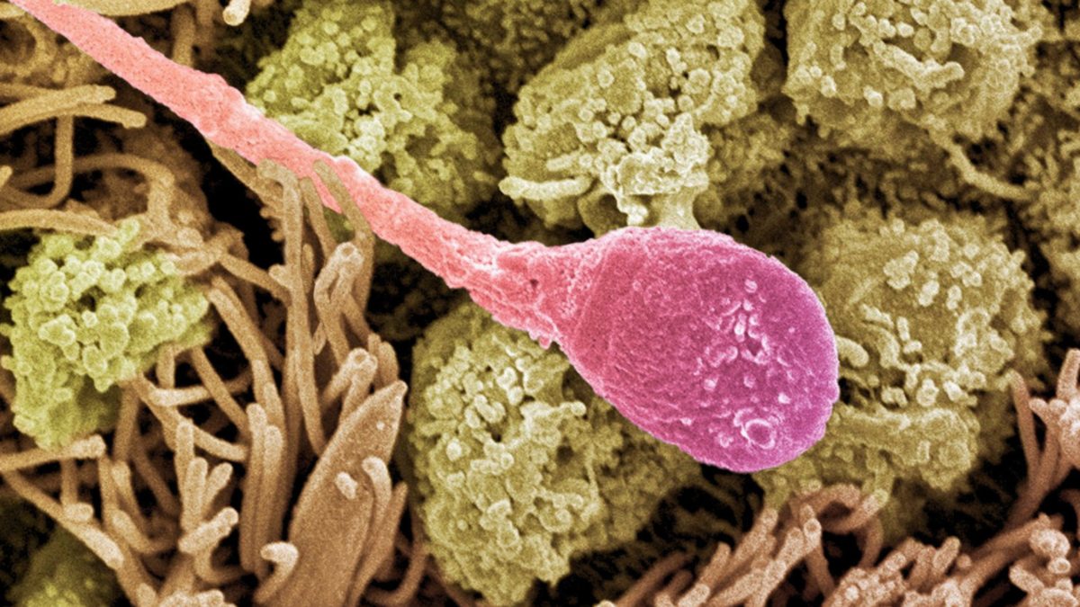
Хулганад хийсэн туршилтаар амилоид бүтэцтэй уургийн ширхэг РНХ-г үрийн эсээр дамжуулан үр хөврөлд хүргэж болохыг харуулжээ.
Эрдэмтэд үрийн шингэн дэх жижиг уургийн ширхэгүүд эргэн тойрны шингэнээс РНХ-г өөртөө холбон, үрийн эсэд нэвтрүүлж болохыг тогтоожээ. Molecular Cell сэтгүүлд нийтлэгдсэн судалгаанд Колумбын их сургуулийн Zuckerman Institute-ийн судлаачид эдгээр ширхэгийг судалсан байна. Ширхэгүүд амилоид бүтэцтэй боловч Альцгеймерийн өвчний үед хуримтлагддаг уургаас өөр уургийн хэсгээс бүрддэг.
Судлаачид түрүү булчирхайн хүчиллэг фосфатаза (PAP) ферментийн жижиг хэсэг болон РНХ-г лабораторид хольж үзэхэд эерэг цэнэгтэй уураг, сөрөг цэнэгтэй РНХ хоорондоо холбогдон дусал үүсгэж, улмаар РНХ агуулсан ширхэг болжээ. Хүний үрийн шингэнд амилоид ширхэгтэй хамт жижиг РНХ, тэр дундаа уураг нийлэгжүүлэхэд оролцдог тээвэрлэгч РНХ-ийн тасархай байгааг мөн илрүүлсэн байна. Үхрийн үрийн эсэд алтны жижиг хэсгээр тэмдэглэсэн ширхэгийг ажиглахад тэдгээр нь эсийн дотор нэвтэрсэн бөгөөд нэмэлт туршилтаар хүний болон хулганы үрийн эс ч ширхэгийг авч байжээ.
Хулганын үрийн эс, өндгөн эсийг ашигласан лабораторийн үр тогтоох туршилтаар PAP-ийн ширхэгээр боловсруулсан үрийн эсээс үүссэн хоёр эсийн шатны үр хөврөлд хүний уургийн хэсгүүд илэрсэн байна. Изотопоор тэмдэглэсэн РНХ-ийг ширхэгт агуулж туршихад ширхэгтэй нөхцөлд үр хөврөлд илүү их РНХ хүрчээ; ногоон гэрэл ялгаруулдаг уургийн заавар бүхий РНХ ашигласан туршилтаар уг уургийн хэмжээ ширхэггүй нөхцөлөөс дор хаяж 10 дахин их байв. Лабораторийн тусдаа туршилтаар кластерин уураг РНХ-г ширхэгээс салгах явцыг түргэсгэсэн ч үр хөврөл доторх энэ үйл явцад ямар үүрэгтэй нь батлагдаагүй байна.
Судалгаа нь үрийн шингэнээс РНХ үрийн эсэд, цаашлаад үр хөврөлд хүрэх боломжит замыг харуулж, РНХ хүргэх арга боловсруулахад чиглэл өгч болох юм. Гэхдээ үр хөврөлд хүргэх туршилтууд хулгана, лабораторид гарган авсан ширхэг болон нэмсэн РНХ-д тулгуурласан бөгөөд хүний үр хөврөл, байгалийн үр тогтолтыг судлаагүй. Байгалиасаа үүсдэг ширхэгүүд өөрийн РНХ-ийн хамт үр хөврөлд нөлөөлдөг эсэх, мөн үр хөврөлд хүрсний дараа ширхэгүүдэд юу болдог нь тодорхойгүй; ширхэг шингэсэн хэмжээг тооцсон дүн ч ашигласан хэмжилтийн аргаас хамааран зөрүүтэй байжээ.
Дэлгэрэнгүйг эх сурвалжаас харах
↓Эх сурвалжийг нээх ↓
A sperm cell’s cargo includesmore than a father’s DNA. RNA molecules that can contribute to early embryonic development also make the journey. Some of those molecules begin their trip outside the sperm.
Researchers have found that tiny protein fibers in semen can help RNA in the surrounding fluid enter sperm cells.
In a new study published in Molecular Cell, the team showed that these fibers can bind RNA, enter sperm, and help deliver that cargo to mouse embryos after fertilization.
The fibers have an amyloid structure, best known for its association with Alzheimer’s disease.
Structural biologist Anthony Fitzpatrick and physiologist William Mallen at Columbia University’s Zuckerman Institute, along with their colleagues, investigated how these fibers might help RNA enter sperm.
“This is wild biology,” Fitzpatrick says.
“Amyloids are most often thought of as a hallmark of Alzheimer’s, Parkinson’s and other neurodegenerative diseases in the brain, but they also have functional roles throughout the body, and keep turning up in unexpected places.”
Amyloid describes the arrangement proteins form when they assemble into fibers. Those studied here are built from a fragment of a protein found in semen, different to the proteins that accumulate in Alzheimer’s.
In semen, they form outside cells in healthy men and can be taken apart again.
Some RNA molecules carry instructions for making proteins, while others help regulate gene activity, giving this cargo a potential role in early development.
The researchers focused on a small fragment of an enzyme produced by the prostate, called prostatic acid phosphatase (PAP). The fragment carried a positive electrical charge, while RNA carried a negative charge.
When mixed in the laboratory, they bound together and formed tiny droplets, which developed into fibers containing RNA.
On their own, the PAP fragments stayed in solution. Adding RNA brought them together and sped up their assembly into fibers. That was the first surprise: RNA helped assemble its own carrier.
“The cargo is helping build the vehicle that carries it,” Fitzpatrick told ScienceAlert.
In human seminal fluid, the team also found small RNA molecules in a fraction containing amyloid fibers. Many identifiable sequences were fragments of transfer RNA, molecules normally involved in assembling proteins.
Finding RNA alongside the fibers didn’t show whether the fibers could enter sperm. Under some imaging conditions, a fiber stuck to a cell’s surface can look much like one sitting within it.
Tiny gold particles attached to laboratory-made fibers allowed the researchers to track them. They used bull sperm for detailed imaging because the cells are relatively large.
A technique that builds three-dimensional images of frozen cells, called cryo-electron tomography, revealed labeled fibers inside sperm.
“Seeing those amyloid fibers inside sperm cells was just so extraordinary to me,” says Mallen.
“There were hints of this happening earlier in our other experiments, but actually seeing this totally blew us away.”
Additional electron microscopy and live-cell imaging supported the finding. Separate experiments showed that human and mouse sperm could also take up the fibers.
“The second surprise was watching sperm take those fibrils up whole and move them deep inside the cell, along the tail, through the midpiece and up into the head,” Fitzpatrick said.
The journey continued in laboratory fertilization experiments using mouse sperm and eggs.
Before fertilization, the team exposed the sperm to fibers made from human PAP fragments. Those human protein fragments also turned up in the resulting two-cell mouse embryos.
To trace the RNA cargo, the researchers loaded the fibers with RNA containing heavier versions of carbon and nitrogen atoms. A technique that distinguishes molecules by mass allowed them to track these labels. More labeled RNA reached the embryos when the fibers were present.
The team tested whether the delivered RNA could function by using RNA that carried instructions for a protein that emits green light. This protein served as a marker showing that the instructions had been used.
Embryos formed from sperm given RNA with the fibers contained at least ten times as much of the protein as embryos formed from sperm given the same RNA without fibers.
Separate laboratory tests showed that a protein called clusterin accelerated RNA’s release from the fibers. Clusterin was also present in the embryos, although its role in releasing RNA inside them remains to be confirmed.
The findings reveal a route by which RNA can travel from seminal fluid into sperm and then into an embryo. Understanding that route could also help researchers design new tools for delivering RNA.
“There are metabolic disorders such as insulin resistance that recent papers have shown can be passed down the paternal line in mice, and it’s thought RNA is how,” Mallen explains.
“By learning how disorders based on epigenetics work, we can come closer to how to possibly treat or prevent them.”
The embryo delivery experiments used mice, laboratory-made fibers, and added RNA. Fitzpatrick said the team hasn’t yet shown whether naturally occurring fibers, together with their usual RNA cargo, change anything in an embryo.
No human embryos or natural conceptions were tested, and uptake estimates varied between measurement methods.
What happens to the fibers after they reach an embryo remains unclear.
“We do not know what becomes of them in the embryo, whether they are cleared, left in place, or seed further aggregation,” Fitzpatrick said.
The research has been published in Molecular Cell.
This article was fact-checked by Rebecca Dyer and edited by Rebecca Dyer. While we pride ourselves on our process, we are only human. If you spot a mistake, please let us know.