Хиймэл оюун ухааны тусламжтайгаар эсийн доторх уурагт бүтцийн хамаарал бүхий вирусэн хувилбарыг анх удаа лабораторийн нөхцөлд бүтээжээ.
Эсийн дотор байрлах, уургаас бүрдсэн хөндий савхан хэлбэртэй “vault” буюу агуулах хэмээх бүтцийг 1986 онд Леонард Ром, Нэнси Кедерша нар анх нээснээс хойш түүний үүрэг зориулалт нь шинжлэх ухааны томоохон таавар хэвээр байсаар ирсэн. Эдгээр бүтэц нь рибосомоос гурав дахин их жинтэй, сүм хийдийн таазны хэлбэрийг санагдуулам бүтэцтэй боловч эсийн үйл ажиллагаанд яг ямар үүрэг гүйцэтгэдэг нь тодорхойгүй байна. Судлаачид эдгээр бүтцийг эсээс бүрэн устгасан туршилтын хулганууд хэвийн хөгжиж, амьд үлдэж байсан нь уг бүтцийн ач холбогдлыг улам бүр эргэлзээтэй болгожээ.
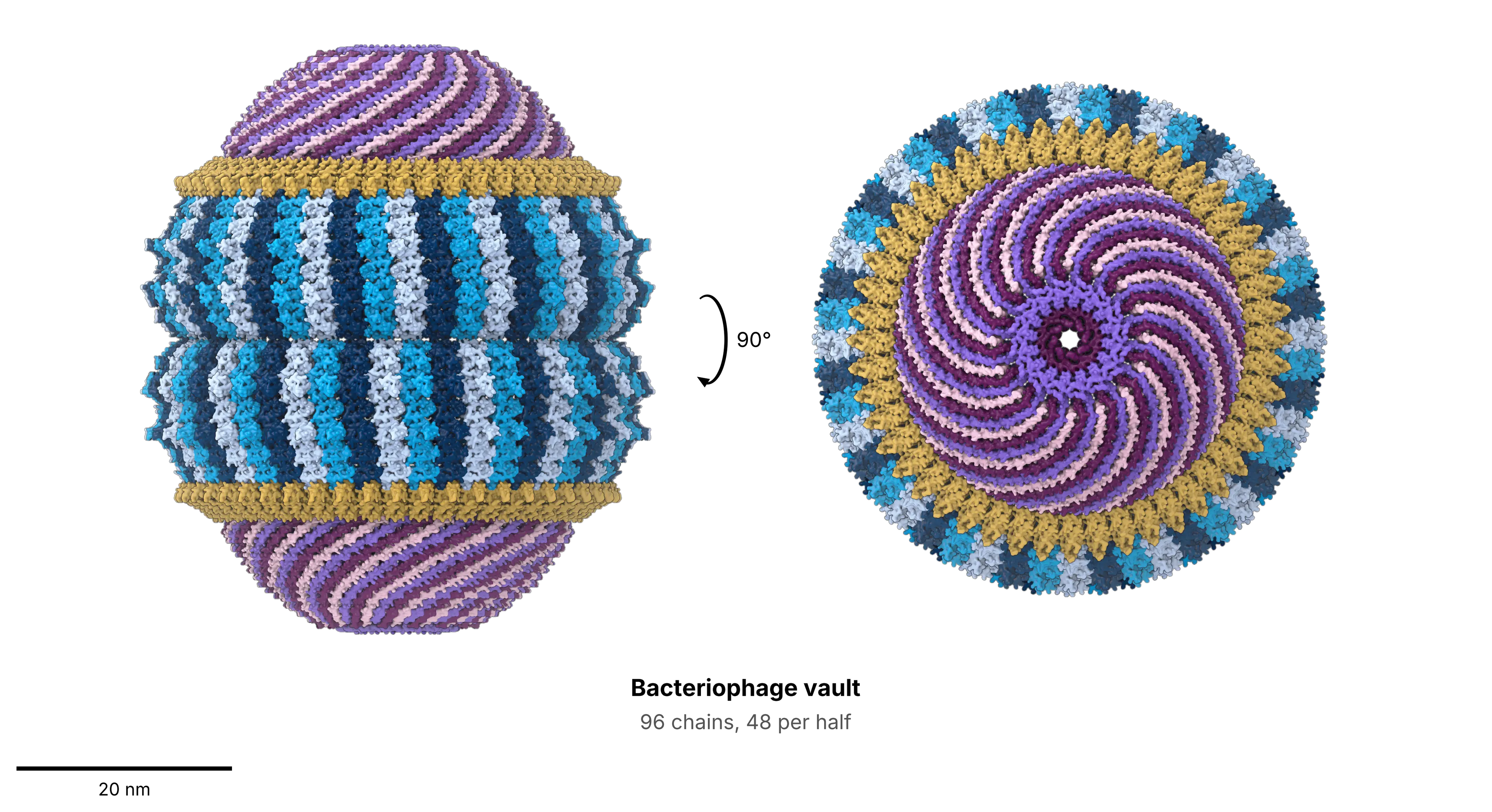
Anthropic компанийн судлаачид Claude загварыг ашиглан 15 тэрбум орчим уургийн дарааллыг шинжилсний дүнд бактери болон бактерийг халдварладаг вирусийн төрөлд агуулахтай төстэй уургууд байгааг илрүүлжээ. Эрдэмтэд 44 нэр дэвшигч уургийг туршиж үзсэний эцэст L05 хэмээх бактериофагийн уургийг ашиглан лабораторийн нөхцөлд 3D бүтцийг нь гарган авч чадсан байна. Энэхүү вирусийн агуулах нь хүний эсийн агуулахтай харьцуулахад илүү том эзэлхүүнтэй бөгөөд 96 уургийн нэгдлээс бүрддэг болох нь крио-электрон микроскопоор хийсэн судалгаагаар тогтоогджээ.
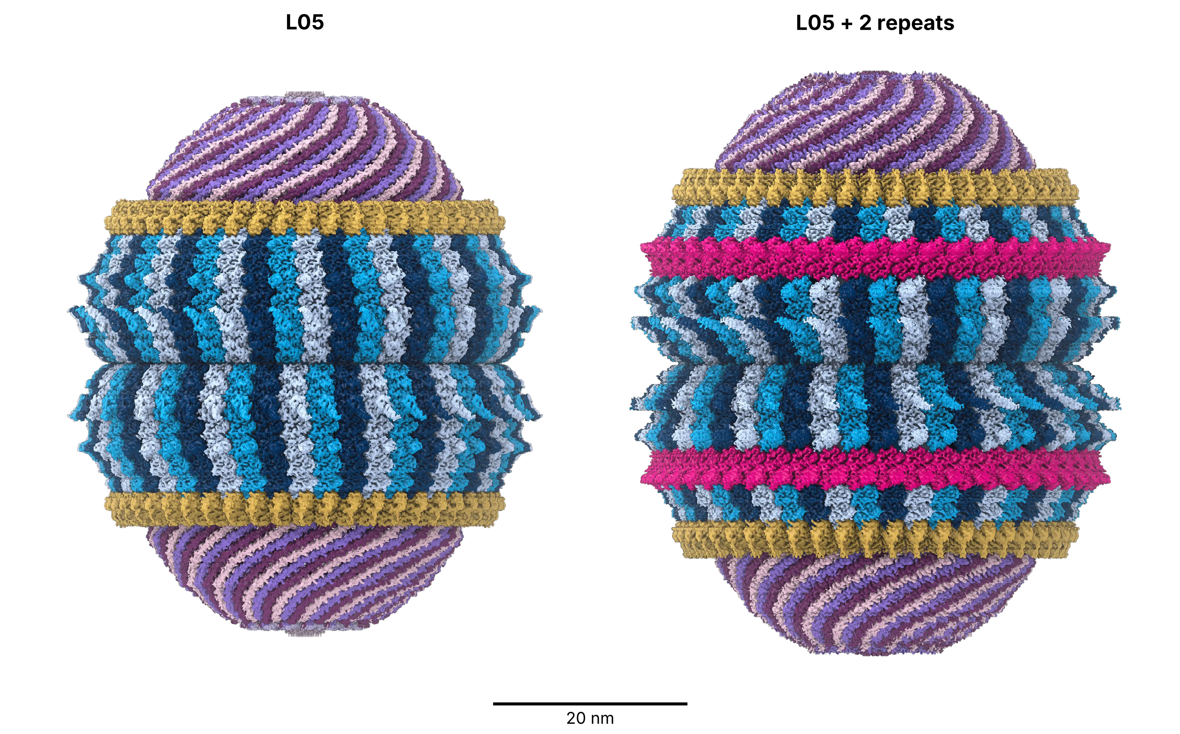
Туршилтын явцад судлаачид уг бүтцийн ханыг өргөтгөн, хүний эсийнхтэй ижил есөн сегменттэй болгож өөрчлөхөд агуулах нь амжилттай угсрагдаж, 20 хувиар илүү багтаамжтай болсон байна. Энэхүү ололт нь эсийн агуулахын бүтэц хувьслын явцад сегментүүдийн хувирлаар өөрчлөгдсөн байж болзошгүйг харуулж буй бөгөөд цаашид эм болон фермент зөөвөрлөх биотехнологийн зориулалтаар ашиглах боломжтойг сануулж байна.
Гэсэн хэдий ч эдгээр бүтэц нь байгалийн жамаар бактери дотор хэрхэн үүсдэг, ямар үүрэгтэй болох нь одоогоор тодорхойгүй байгаа юм. Судлаачид L05 агуулахын бүтцээс химийн бүлэг зөөвөрлөгч коэнзим А-тай төстэй молекул илрүүлсэн нь уг бүтцийн үүргийг тайлах нэгэн сэжүүр байж болох ч нарийвчилсан судалгаа шаардлагатай хэвээр байна.
Дэлгэрэнгүйг эх сурвалжаас харах
↓Эх сурвалжийг нээх ↓
Almost every cell in your body contains tiny, hollow barrels built from protein. These structures are enormous by molecular standards, weighing roughly three times as much as a ribosome. Scientists discovered them 40 years ago, named them, and have spent much of the time since arguing over what they might be doing.
Nobody has a definitive answer.
Now, researchers have tracked down close relatives of these mysterious structures in an unexpected place: viruses that infect bacteria. One of the viral versions has been reconstructed in the laboratory, and it turns out to be bigger than the structures found in humans.
Named After a Cathedral Ceiling
Back in 1986, Leonard Rome and Nancy Kedersha were examining an unfamiliar protein structure under an electron microscope. Its ribbed shell looked rather like the vaulted ceiling of a cathedral. They called the structures vault particles, and the name stayed.
A mammalian vault contains 78 copies of the same protein, assembled into two matching halves that meet at the center. Vaults occur throughout much of the eukaryotic world, from slime molds to humans. Their evolutionary history stretches back hundreds of millions of years, which makes their stubbornly unknown function all the more puzzling.
Over the years, researchers have suggested several possible jobs. Perhaps vaults carry molecules between the nucleus and the rest of the cell. Perhaps they help cells withstand toxic substances, including cancer drugs, or participate in immune defense. So far, none of those explanations has been firmly established.
Then came a particularly awkward finding. Scientists engineered mice that lacked vaults entirely, expecting the loss might reveal what these structures were needed for. Instead, the mice developed normally and survived.
Whatever vaults do, they do not appear essential for a mouse living under ordinary laboratory conditions. Making matters worse for researchers, several of biology’s favorite experimental organisms, including baker’s yeast, fruit flies and nematode worms, lack vaults altogether.
The researchers needed another place to look.
A Search Through 15 Billion Proteins
That search led to the biology research lab at Anthropic, the AI company, where researchers used Claude models throughout the project. Earlier bioinformatics work had turned up distant relatives of the vault protein in bacteria and in viruses that infect bacteria. But a sequence resemblance was one thing; showing that those proteins could actually assemble into vaults was another.
The team combed through a database containing approximately 15 billion predicted proteins. Vault-like proteins appeared across 29 major bacterial groups and four groups of archaea, although they occurred in fewer than 1% of species with sequenced genomes.
From that enormous search, the scientists selected 44 candidates for experimental testing. Fifteen could be produced in living E. coli cells. Getting the proteins to assemble was only part of the challenge, however. The researchers still had to extract intact particles from the bacteria without wrecking them.
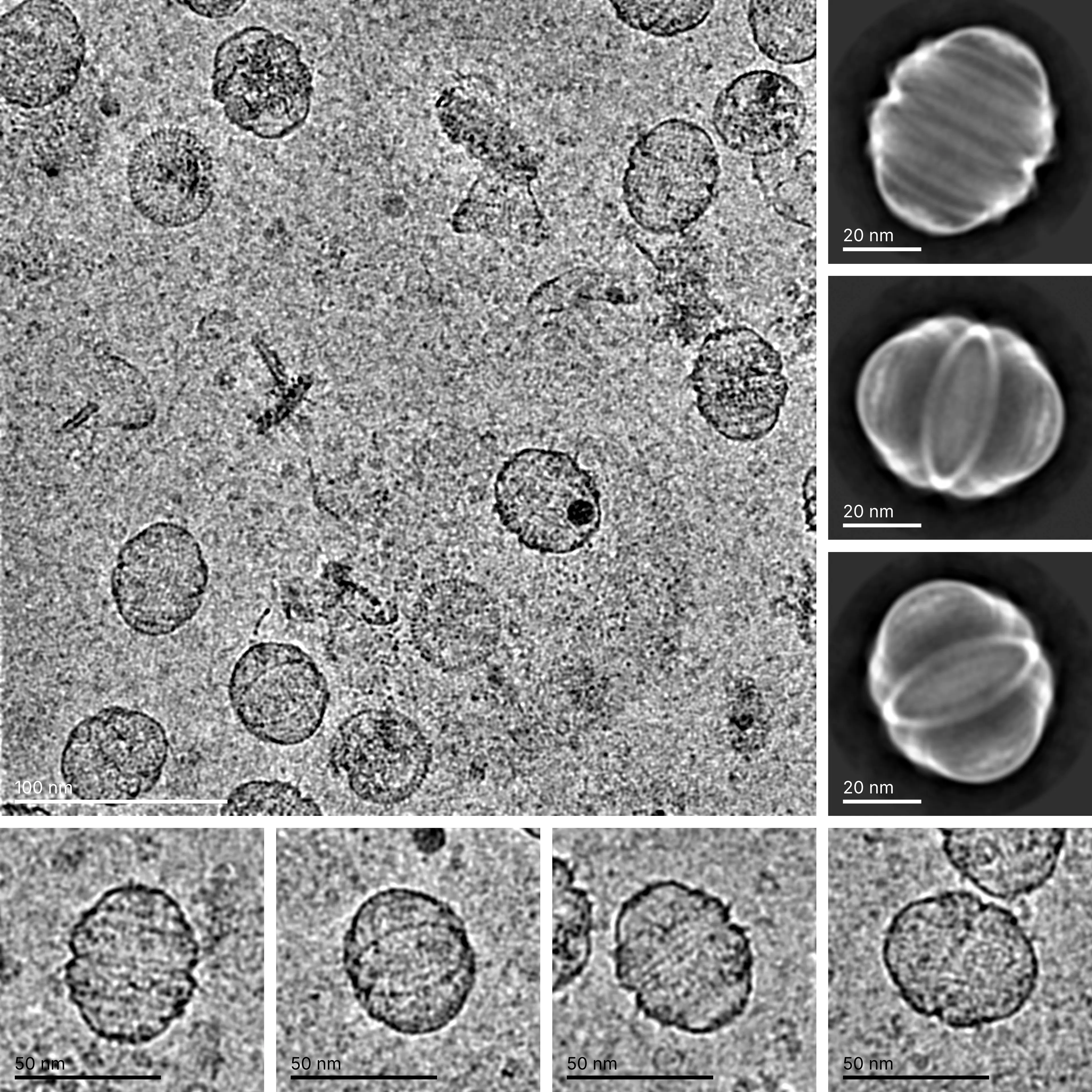
Traditionally, scientists isolate animal-cell vaults by spinning samples through a gradient of increasingly concentrated sugar. The procedure can take about three days. Rather than follow that route, the Anthropic team developed a method using a column packed with porous beads, allowing the largest particles to emerge first from the broken-up cells.
After optimization, the process took roughly 90 minutes, from cells to microscope. Electron microscopy revealed vault-shaped assemblies from seven of the 15 proteins tested, and four were subsequently confirmed as genuine vaults.
Two belonged to bacteria. The other two had a different origin.
A Vault Built by a Virus
Those two proteins were encoded by bacteriophages, viruses that infect bacteria and commandeer their cellular machinery to manufacture viral components. The finding meant that a bacterial cell could, at least in principle, construct vaults according to instructions supplied by an invading virus.
For a closer look, the researchers selected a phage protein called L05. Using cryo-electron microscopy, which involves flash-freezing large numbers of particles and combining their images into a three-dimensional reconstruction, they obtained the first 3D structure of a vault from a prokaryotic cell.
The viral vault was noticeably different from its human counterpart. Instead of 78 protein copies, it contained 96, with 48 forming each half. Its overall shape was shorter and broader, measuring approximately 54 nanometers in length and 45 nanometers across, compared with 67 by 40 nanometers for a human vault. Despite its shorter length, the rounder viral structure enclosed roughly 25% more volume.
There were differences in the shell, too. Human vault walls contain nine repeating segments stacked along their length, whereas the phage version has seven. Its end caps are flatter, and the structure ranks among the largest protein shells identified in prokaryotes.
How those caps close is particularly interesting. Each half begins with a wall 48 protein copies around, but the wall must narrow toward the tip. It does so through three successive steps, forming something like a tiered dome.
Within each group of three neighboring protein copies, one ends at the first step, another reaches the second, and the last continues all the way to the pole. The proteins are chemically identical, yet each adopts a different shape.
Building a Bigger Vault
Once the researchers understood the architecture, they began changing it. They engineered 15 modified versions of the L05 protein, concentrating largely on the regions where adjacent copies make contact. A few single changes near the cap were enough to prevent closure, leaving open, rosette-shaped assemblies instead of complete barrels.
That gave the team a clearer picture of which regions were responsible for sealing the shell.
Next came an attempt to increase its capacity. The scientists duplicated two of the seven repeating wall segments, producing a protein with nine segments, the same number present in human vaults. To their surprise, the redesigned proteins assembled successfully into larger particles.
The modified vaults offered about 20% more internal space. Such a straightforward enlargement suggests that vaults might have changed size during evolution through duplication of existing structural segments. It also makes them intriguing candidates for biotechnology, where hollow protein shells could potentially serve as containers for delivering drugs or enzymes.
The experiments have not, however, settled the question that has followed vaults since their discovery. No biological function has been established, and the team has so far produced these structures only in E. coli. Whether the same particles assemble naturally in the bacteria and phages carrying the relevant genes remains to be demonstrated.
Rome, who helped discover vaults in 1986 and served as a paid consultant to the Anthropic team, acknowledged the problem. “Figuring out the function of vaults is still enormously challenging,” he said. Given how widely the newly identified relatives are scattered, he expects “a complicated function and an interesting ride.”
One possible lead is already visible in the structural data. Partway along each of the 96 protein copies sits a small pocket, and the cryo-electron microscopy map shows something lodged inside it. The researchers have not identified the molecule, although its shape resembles coenzyme A, a compound cells commonly use to transfer chemical groups.
Enjoyed this article? Subscribe to our free newsletter for engaging stories, exclusive content, and the latest news.