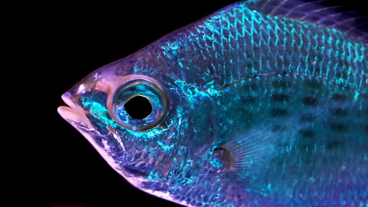
Японы Хирошима их сургуулийн инженер Масаказу Ивасака Япон болон Солонгосын эрэг орчмын гүехэн усанд амьдардаг Hypoatherina tsurugae хэмээх загасны арьсны бүтцийг судалжээ. Эдгээр загасны арьсанд гуанины талстаар дүүрсэн “иридофор” хэмээх эсүүд байдаг бөгөөд тэдгээр нь гэрлийг өөр өөр өнцгөөр ойлгон өнгө үүсгэдэг байна.
Судалгааны явцад эдгээр эс нь зөвхөн гэрэл ойлгоод зогсохгүй, орчны гэрэлтүүлгийн өөрчлөлтөөс хамааран идэвхтэй өөрчлөгддөг болохыг ажиглажээ. Судлаач загасыг LED гэрлээр гэрэлтүүлэх үед иридофор эсүүд “гэрэлт” төлөвөөс гэрэл унтраахад шууд “харанхуй” төлөвт шилжиж байв. Энэхүү хариу үйлдэл нь ялангуяа цэнхэр өнгийн гэрэлд хамгийн мэдрэг байсан бөгөөд гэрэл унтрааснаас хойш 10 орчим секундын дотор эсүүд анхны байдалдаа эргэн орж байжээ.
Энэхүү хурдан бөгөөд уян хатан хариу үйлдэл нь загасны арьсанд гэрэл мэдрэх чадвар байгааг илтгэж байна. Масаказу Ивасакагийн таамаглаж буйгаар, энэхүү үйл явцад нүдний торлог бүрхэвчид байдаг гэрэл мэдрэгч уураг болох опсинууд эсвэл мэдрэлийн системийн оролцоо байж болзошгүй аж. Одоогоор уг судалгаа нь хөндлөнгийн хяналт (peer review)-д ороогүй урьдчилсан хэвлэл бөгөөд цаашид энэхүү үзэгдлийн молекулын түвшний механизмыг нарийвчлан судлах шаардлагатай байна.
Дэлгэрэнгүйг эх сурвалжаас харах
↓Эх сурвалжийг нээх ↓
A glittering school of silverside fish may remind you of a disco ball, a dazzling display made up of many tiny mirrors.
But emerging research suggests the skin of these fish is capable of far more than just reflecting light.
Masakazu Iwasaka is an interdisciplinary engineer at Hiroshima University in Japan who has been researching the interplay of light across the bodies of fish for decades.
In a new preprint paper, which has not yet been peer-reviewed or formally published, Iwasaka says he has found specialized pores in fish skin actually reacting to light, not just reflecting it.
He observed this remarkable phenomenon in cobalt silverside fish Hypoatherina tsurugae, which are found in relatively shallow ocean waters around Japan and Korea.
Embedded within their skin, these fish have special kinds of cells known as iridophore cells, densely packed with crystals of guanine.
These crystals reflect light at different angles to produce the appearance of color, not via pigment, as our skin cells do with melanin, but by their structural arrangement, which bends and reflects light at different wavelengths.
In previous studies on these fish, Iwasaka recorded iridiphore spots along the animals’ dorsal trunk scales that exhibited “rapid and repetitive light reflection at frequencies of several hertz, independent of body motion”.
These spots switch between three states, which Iwasaka rather poetically named ‘static bright’, ‘dynamically twinkling’, and ‘dark’.
He suspected these states may change depending on environmental light levels, and designed this new study to test that theory.
Iwasaka conducted his experiments on 22 fish, caught from the wild and kept in a standard lab aquarium.
To get a microscopic look at their skin under changing light conditions, some fish were briefly anesthetized, examined with a microscope-lens camera, and then returned to the main aquarium.
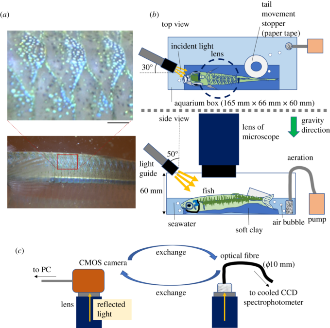
First, he aimed a white LED at the fish, which was the same kind of light illuminating the room and the aquarium generally.
The iridophores remained in their ‘static bright’ state – until the direct light was switched off, with the iridophores quickly transitioning to ‘dark’ mode, a bit like how our pupils would change if someone turned off the lights.
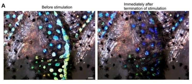
In this ‘dark’ state, the guanine crystals stopped reflecting light with the same intensity, as if they had been shifted slightly.
But before long, they adjusted back to the ambient white light of the room, returning to ‘static bright’ mode.
Then, Iwasaka repeated this experiment, testing how the iridophores reacted to blue, green, and red LED lights, as well as a blue laser, and two kinds of green lasers.

“Spectral analysis revealed that this quenching response was most sensitive to blue light compared with green and red illumination,” Iwasaka describes in his paper.
In these experiments, he says, iridophores returned to a twinkling state within about 10 seconds after the direct light source was cut.
“The speed and reversibility of this response suggest that mechanisms beyond slow intracellular structural rearrangements are involved and raise the possibility of neural modulation in addition to intrinsic photoreceptive processes,” Iwasaka writes.
Related: The Prettiest Sea Slugs Use Crystals in Their Skin as ‘Pixels’ of Color
He suspects opsins – a group of light-sensitive proteins known best for their role in our retinas – may be involved.
But that theory is beyond the scope of the current study, which stopped short of probing the molecular mechanisms behind the fish skin’s light reactivity.
Please note this study has not yet undergone the rigorous process of scientific peer review or formal publication. This means it could still contain errors, omissions, and is not yet endorsed by the scientific community.
It was uploaded to the preprint server bioRxiv.
This article was fact-checked by Rachel Garner and edited by Peter Dockrill. While we pride ourselves on our process, we are only human. If you spot a mistake, please let us know.