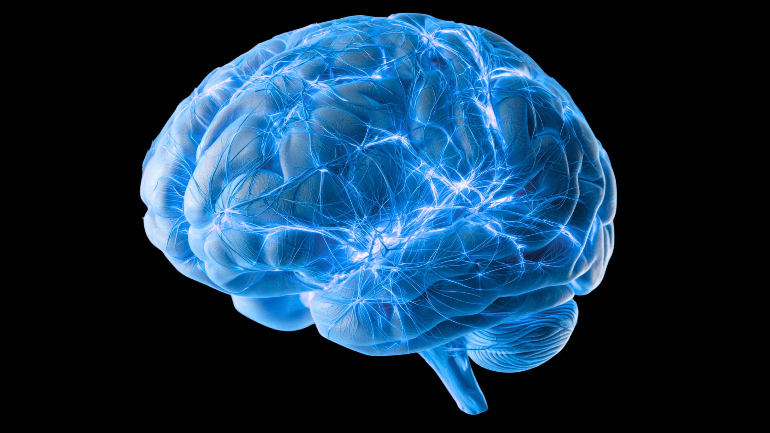
Эрдэмтэд тархинд хуримтлагдсан хортой уургийг гадагшлуулахад оролцдог өмнө нь бүртгэгдээгүй жижиг сувгуудын сүлжээг олж тогтоожээ.
University College London-ийн судлаачид хулганы тархины эдэд хийсэн туршилтаар флюресцент тэмдэглэгээтэй уургууд нь тархины гадаргуу руу чиглэсэн нарийн сувгуудаар дамжин гадагшилдаг болохыг илрүүлжээ. Энэхүү сувгууд нь ойролцоогоор 2 микрометрийн диаметртэй бөгөөд фибробласт торлог эсүүдээс бүрдсэн сүлжээ үүсгэдэг байна. Судлаачид мөн мэс заслын аргаар авсан хүний тархины эдэд ижил төстэй бүтэц байгааг ажигласан нь хүн дээр ч мөн ижил төстэй гадагшлуулах зам байх магадлалтайг харуулж байна.
Альцгеймерийн өвчний үед амилоид бета болон тау уураг тархинд хуримтлагддаг нь мэдрэлийн эсүүдэд сөргөөр нөлөөлдөг. Энэхүү судалгаа нь хортой уургууд хэрхэн гадагшилдаг, яагаад хуримтлагддаг болохыг ойлгох, цаашлаад Альцгеймерийн өвчний эмчилгээний шинэ гарцыг хайхад чухал ач холбогдолтой юм. Судлаач Дэвид Атвеллийн тайлбарласнаар, энэхүү сувгууд нь хүний биеийн тунгалгийн зангилаатай төстэй үүрэг гүйцэтгэдэг байж болзошгүй аж.
Гэсэн хэдий ч судалгааны үр дүн нь одоогоор хянан магадлагаанд ороогүй (preprint) бөгөөд амьд хүний тархинд эдгээр сувгууд хэрхэн үйлчилдэг, уургийг гадагшлуулах хөдөлгөх хүч нь юу болох нь тодорхойгүй байна. Судлаачид уургийн шилжилт хөдөлгөөнийг юу өдөөж байгаа болон энэ үйл явц удаашрах нь өвчлөлийн эхлэлд нөлөөлдөг эсэхийг ирээдүйд нарийвчлан судлах шаардлагатай гэж үзэж байна. Одоогийн байдлаар энэхүү судалгаа нь эмчилгээний арга барилыг батлаагүй бөгөөд зөвхөн тархины цэвэрлэгээний системийн бүтцийн талаарх шинэ мэдээллийг өгч байна.
Дэлгэрэнгүйг эх сурвалжаас харах
↓Эх сурвалжийг нээх ↓
You read a sentence, remember a familiar face, then turn to another thought. Behind all of that activity, your brain also has a housekeeping problem: what to do with proteins that can become harmful when they accumulate.
Those proteins need an exit. But tracing their journey out of the brain has proved surprisingly difficult.
Now, researchers at University College London have identified a previously undocumented network of tiny channels that could fill in part of the map.
In mice, fluorescently labeled proteins entered these channels and traveled towards the brain’s surface. Similar structures appeared in human brain tissue removed during surgery, raising the possibility that we share this clearance route.
The study is a preprint that has not undergone peer review, and the channels’ function in living humans remains to be established.
“The pathways by which toxic proteins are removed from the brain are poorly understood,” neuroscientist David Attwell told ScienceAlert.
This knowledge gap is a problem for researching Alzheimer’s disease, which involves the accumulation of amyloid beta and abnormal tau proteins. Understanding why they build up requires investigating both their production and their removal.
Scientists have proposed different routes out. One follows the walls of arterioles, small vessels supplying brain tissue with blood. Another follows the spaces around venules, small vessels that carry blood away, and features in explanations of the glymphatic system.
Earlier research has explored fluid movement along the brain’s blood vessels. The new study identifies a specific cellular structure that may help explain how proteins travel through two proposed exit routes.
“We have now shown that, in both these locations, proteins are in fact removed through tiny tubes that appear to be similar to the tubes present in lymph nodes outside the brain (e.g. in the armpit or neck),” Attwell explained.
The channels measure approximately 2 micrometers across and form an interconnected mesh. They extend along blood vessels into the membranes covering the brain and along nerves leaving it.
The findings suggest they are formed by fibroblastic reticular cells. Related cells build networks that carry fluid and dissolved substances through lymph nodes.
To follow what moved through the channels, the researchers made tau and amyloid beta visible using fluorescent labels.
In mouse experiments, the labeled proteins built up inside the channels as they moved. In some experiments, tracers reached channels at the upper surface within 7.5 minutes of administration.
Tau traveled through both arteriole walls and channels around venules. Measurements suggested roughly similar amounts passed through each route, supporting the possibility that they belong to a shared network.
Other clearance mechanisms may operate alongside it. Some proteins can be broken down within brain tissue or pass across blood vessel cells into the bloodstream.
The researchers also investigated whether the network handled proteins produced inside the brain, rather than only substances introduced during experiments.
In mice modeling aspects of Alzheimer’s disease, they found the animals’ own amyloid beta within the channels. This supports a role in transporting naturally produced proteins, although it does not establish how much ultimately exits through the network.
The human findings came from five patients undergoing surgery for brain tumors. The team examined living tissue removed during their operations that would otherwise have been discarded.
When fluorescently labeled tau and amyloid beta were applied to the human tissue, the proteins concentrated in channels resembling those in mice. Their dimensions were similar, too.
But tissue maintained outside the body cannot reveal the complete clearance process inside a living person.
“In the future it will be important to demonstrate that these tubes function in the same way in living humans,” Attwell said.
Another mystery concerns what drives the movement. The researchers do not yet know precisely how proteins enter the channels or how fluid travels through them so quickly. Pulsing blood vessels might contribute, but the pumping mechanism remains unresolved.
Understanding that mechanism could help researchers investigate whether impaired transport contributes to disease, and whether changing the flow would make a difference.
“We also need to know whether a slowing of this pumping contributes to initiating Alzheimer’s disease, and whether therapeutically speeding the pumping could help treat the disease,” Attwell said.
The study did not test a treatment or demonstrate improvements in memory. Those possibilities require further research.
For now, scientists have a newly identified structure to investigate. Following proteins through these tiny channels could help answer a much larger question: when potentially harmful substances accumulate in the brain, where does their journey out go wrong?
A preprint of the study is available on bioRxiv.
This article was fact-checked by Rebecca Dyer and edited by Rebecca Dyer. While we pride ourselves on our process, we are only human. If you spot a mistake, please let us know.


Тархинд хуримтлагддаг хортой уургуудыг гадагшлуулах шинэ сувгийн сүлжээг илрүүлж байгаа нь маш сонирхолтой санагдаж байна. Энэ судалгаа нь хүний өвчин үүсэх механизмийг ойлгоход ямар нөлөөтэй бол гэж бодож байна вэ?