Дархлааны тогтолцооны CD4+ T эсүүд нь өмнө нь тооцоолж байснаас өөрөөр хавдрын эсүүдийг устгах чадвартай болохыг судалгаагаар тогтоожээ.
Мичиганы их сургууль болон Baylor College of Medicine-ийн судлаачдын баг хавдрын эсүүд дархлааны тогтолцооноос зугтахын тулд өөрсдийн таних тэмдэг болох MHC I молекулыг нуудаг болохыг ажиглажээ. Уламжлалт ойлголтоор MHC I молекул байхгүй тохиолдолд CD8+ T эсүүд хавдрыг таньж чаддаггүй байсан бол шинэ судалгаагаар энэ нөхцөлд CD4+ T эсүүд хавдрын эсүүдийг устгах үүрэг гүйцэтгэдэг болохыг илрүүлсэн байна.
Туршилтын хулгана болон хүний эмнэлзүйн генетик мэдээллийн санд хийсэн судалгаагаар CD4+ T эсүүд нь “ферроптоз” буюу төмрөөс хамааралтай эсийн үхлийн процессыг ашиглан хавдрын эсийг устгаж байгааг тогтоожээ. Мөн энэхүү механизм нь шилжүүлэн суулгасан үүдэл эс донорын эд эрхтнийг гэмтээдэг “graft-versus-host disease” (GVHD) өвчний үед гэдэсний эсүүдийг устгахад нөлөөлдөг болохыг судлаачид тэмдэглэв.
Immunologist Pavan Reddy-ийн мэдээлснээр, энэхүү нээлт нь хавдрын дархлаа эмчилгээний шинэ стратеги боловсруулах болон дархлааны хариу урвалыг удирдах боломжийг бүрдүүлж болзошгүй юм. Гэсэн хэдий ч энэхүү үйл явц нь өөр төрлийн хавдар болон аутоиммун өвчний үед хэрхэн нөлөөлөхийг цаашид нарийвчлан судлах шаардлагатай байна.
Судалгааны үр дүнг Nature Immunology сэтгүүлд нийтэлжээ.
Дэлгэрэнгүйг эх сурвалжаас харах
↓Эх сурвалжийг нээх ↓
In the army of defenders that is the body’s immune system, each cell has specific targets for which it surveils.
As soon as it spots a marker of a foreign invader or infected cell, it launches an attack or recruits other cells to help, rallying an immune response.
But researchers have just discovered something unexpected about how the immune system fights cancer, something which upends our long-held view of which targets different immune cells attack.
At the crux of the discovery is a sneaky trick that cancer cells sometimes use to evade detection by our natural defenses: They essentially hide their ID badges, known as major histocompatibility complexes or MHCs.
That deception helps cancer cells evade detection from CD8+ T cells, the primary, destroyer-type immune cells also known as ‘killer’ T cells.
With this new discovery, however, scientists have realized that cancer cells can become more vulnerable to other immune cells previously seen as more of a secondary support squad, CD4+ T cells – or ‘helper’ T cells.
The findings, from researchers at the University of Michigan and the Baylor College of Medicine in the US, challenge decades of immunology research, and could have implications beyond cancer, too.
“Our work, if further validated, will have implications for T cell-mediated immune responses beyond cancer and transplant immunology,” says immunologist Pavan Reddy, from the Baylor College of Medicine.
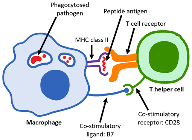
There are two main classes of MHCs. Class I are reported by all nucleated cells (except red blood cells), to tell the immune system who they are and what they’re up to.
Class II are a bit different: They’re expressed specifically by immune system scouts, such as macrophages, as alerts for T cells about potentially harmful material in the body.
Previously, it was thought that cells with suspect MHC I markers were hunted down by CD8+ T cells, while cells flagged by MHC II were cleared by CD4+ T cells.
The new results from tests on experimental mice, validated in genetic databases recorded from treatments on human patients, showed that cancer cells shirking their MHC I badges could still be attacked by CD4+ T cells.
“While pathogens and tumor cells often downregulate MHC I-mediated antigen presentation to escape from immune surveillance, our observations now suggest that this deficiency may paradoxically sensitizes them to CD4+ T cell-mediated elimination,” write the researchers in their published paper.
“Thus, we expand the scope of MHC I from the long-held paradigm in T cell immunity that MHC I exclusively mediates only CD8+ T cell responses.”
The researchers also studied mouse models of graft-versus-host disease (GVHD), a condition where transplanted stem cells from a donor mistakenly attack the body’s healthy tissues.When MHC I badges were absent in these models, CD4+ T cells were still able to kill target intestinal cells, explaining how donor immune cells may harm the gut.
The researchers identified how CD4+ T cells carry out their work in these scenarios: They use a process known as ferroptosis, where cells are killed off by an iron-dependent form of programmed cell death.
“We now identify ferroptosis as a contributor to the severity of gastrointestinal-GVHD,” the researchers write.
“Whether ferroptosis contributes to other target organ damage must be determined in future studies.”
As this is a brand-new discovery, there’s still more to do to confirm it works as the researchers suggest, but further down the line, we might be able to use this knowledge to our advantage in developing cancer treatments.
Immunotherapies – treatments that aim to make the body’s natural immune system more effective – are becoming more widely used for various cancers.
This new discovery could help make immunotherapies more potent, now that we know there is another way to thwart a self-preservation method used by cancer cells.
“This may allow for the development of novel strategies that target MHC class I and CD4+ T cells to leverage the beneficial side of immunity or mitigate unwanted immune responses,” says Reddy.

As the main research here involved mice, the researchers hope to take these findings further with a closer analysis of the biological mechanisms driving CD4+ T cells in clinical studies.
It will also be important to look in more depth at how the T cell tag-team effort applies across different types of cancer.
CD4+ T cells are also known to play a role in other autoimmune conditions, including type 1 diabetes, where immune cells wrongly destroy insulin-producing cells in the pancreas, and celiac disease. So there could be a lot to discover.
Related: A Breakthrough Drug Just Achieved The ‘Impossible’ For Pancreatic Cancer
“Our study provides new insights into the role of MHC I in CD4+ T cell-mediated immunity, unveiling mechanisms distinct from its canonical role in antigen presentation to CD8+ T cells,” write the researchers.
The research has been published in Nature Immunology.