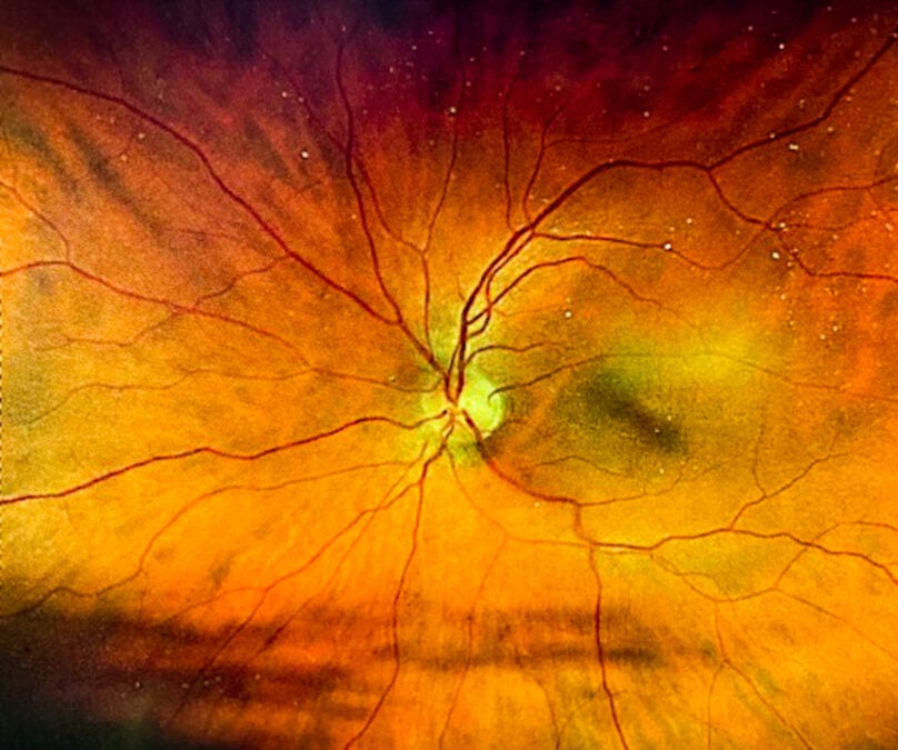
Судлаачид нүдний торлог бүрхэвчийн бүтцийн өөрчлөлтийг шинжлэх замаар Альцгеймерийн өвчин үүсэх эрсдэлийг урьдчилан таамаглах боломжтойг тогтоожээ.
Флоридагийн их сургуулийн биоанагаахын инженер Руогу Фан тэргүүтэй судлаачдын баг 40,000 гаруй UK Biobank-ийн оролцогчийн 62,876 ширхэг торлог бүрхэвчийн зурагт машин сургалтын технологийг ашиглан дүн шинжилгээ хийжээ. Уг загвар нь нас, хүйс, нойрны чанар, согтууруулах ундааны хэрэглээ, сэтгэл гутрал, цусны даралт зэрэг Альцгеймерийн өвчний эрсдэлтэй холбоотой 12 хүчин зүйлийг урьдчилан таамаглахад чиглэсэн байна.
Судалгаагаар торлог бүрхэвч дэх судасны хатуурал, цусны судасны нягтрал буурах болон харааны мэдрэлийн эдийн шингэрэлт зэрэг нь Альцгеймерийн өвчний эрсдэлтэй холбоотой болохыг тогтоожээ. Ялангуяа хожим энэ өвчнөөр оношлогдсон хүмүүсийн торлог дахь жижиг артерийн судаснууд агшсан шинж тэмдэг илэрсэн байна. Энэ нь тархины мэдрэл-судасны үйл ажиллагааны доголдолтой холбоотой байж болох ч одоогоор энэ нь таамаглал төдий бөгөөд цаашид нарийвчлан судлах шаардлагатай гэж эрдэмтэд тэмдэглэжээ.
Руогу Фангийн тайлбарласнаар, Альцгеймерийн өвчин олон арван жилийн турш хөгждөг боловч одоогийн оношилгооны аргууд ихэвчлэн өвчнийг хүндэрсэн үед нь илрүүлдэг байна. Торлог бүрхэвчийн зураглал нь өвчнийг эрт оношлох бус, харин өндөр эрсдэлтэй бүлгийг ялгаж, амьдралын хэв маягаа өөрчлөх болон урьдчилан сэргийлэх арга хэмжээ авах боломжийг олгох “биологийн мэдрэгч”-ийн үүрэг гүйцэтгэх боломжтой.
Энэхүү судалгааны үр дүн нь Journal of Alzheimer’s Disease сэтгүүлд нийтлэгджээ. Торлог бүрхэвчийн зураг нь Альцгеймерээс гадна ясны эрүүл мэнд болон эрт нас барах эрсдэлийг ч харуулж болзошгүй гэсэн нотолгоонууд нэмэгдсээр байна.
Дэлгэрэнгүйг эх сурвалжаас харах
↓Эх сурвалжийг нээх ↓
With every year, more evidence is connecting our eyes to Alzheimer’s disease.
The retina is the light-sensitive tissue at the very back of the eye, and it contains several layers of neurons.
A change here could be one of the most obvious outward signs of a deeper problem, hidden beyond our sight.
Already, scientists have shown that photos of the retina can identify those with active and ongoing cases of Alzheimer’s disease.
Now, some of the same researchers have shown that these photos can help to determine a person’s risk of developing Alzheimer’s, years before diagnosis.
To be clear, these photos can’t actually diagnose Alzheimer’s years prior to symptoms.
What they can do is reveal subtle signatures of factors that may contribute to Alzheimer’s risk.
This adds to emerging evidence that imaging tiny blood vessels and nerves in the eye could provide a ‘heads up’ for those at higher risk of cognitive decline.
“We know that Alzheimer’s disease develops over decades, but most of the diagnostic tools focus on late-stage pathology when it is too late to intervene,” explains biomedical engineer Ruogu Fang from the University of Florida, who led the new study.
“By looking at novel biomarkers, like retinal health, we offer new opportunities to identify patients at risk, offer appropriate tests, and encourage them to develop healthy lifestyles to mitigate their risk.”
The team used machine learning to analyze 62,876 retinal photographs from more than 40,000 UK Biobank participants.
Their AI program was designed to predict 12 factors linked to a higher risk of Alzheimer’s disease, such as sex, smoking, sleep, alcohol use, depression, age, body mass, and blood pressure.
Ultimately, the deep learning models identified several subtle markers associated with the development of Alzheimer’s disease.
Vascular stiffening, reduced blood vessel density, and thinning of the optic nerve, for instance, are signs of retinal aging that are also associated with Alzheimer’s.
Predictive models about who would go on to develop Alzheimer’s relied on demographic, vascular, metabolic, and lifestyle risk factors, as well as retinal images, and their corresponding risk factors.
Patients who would later go on to develop Alzheimer’s, for instance, showed constriction of small arteries and arterioles in their retina, which aligns with the findings of previous research.
This could be a sign of neurovascular dysfunction more broadly, although that explanation remains speculative.
These are just associations that need to be probed further.

“Existing work has largely focused on predicting individual risk factors or disease states in isolation, without systematically examining how retinal structural patterns relate to a broader spectrum of Alzheimer’s disease-related risk domains,” write Fang and colleagues.
Photographs of the retina are already routinely taken for those with diabetes, glaucoma, or cataracts.
If these images contain clues to dementia risk, then they could be really useful data points to track the progression of brain disease.
Some evidence even suggests that the retina could hold clues to our general well-being, extending far beyond the brain.
Studies indicate that this layer of the eye may predict a person’s bone health or even their risk of early death.
Perhaps that is because the retina faithfully reflects signs of some underlying diseases.
Alzheimer’s could be among several other illnesses reflected in the eye.
Related: A Small Quirk in Your Eye Movement Could Indicate Alzheimer’s
“In this sense,” says Fang, “retinal imaging functions less as a surrogate questionnaire and more as an integrated biological sensor of cumulative risk.”
Like the trail of Hansel and Gretel, Alzheimer’s disease may scatter crumbs as it progresses along.
Tracing those clues back to the source could give scientists a whole new understanding of where this disease comes from and why.
The study is published in the Journal of Alzheimer’s Disease.