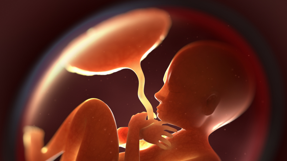
Эрдэмтдийн шинээр боловсруулсан ихэсийн загвар нь микро болон нано хэмжээтэй хуванцар тоосонцор хүний биеийн эсүүдэд хэрхэн нөлөөлдгийг харууллаа.
Vrije Universiteit Amsterdam-ийн судлаач Жеске ван Боксел болон түүний багийнхан Molecular and Cellular Endocrinology сэтгүүлд нийтлүүлсэн судалгаандаа хүний ихэсийн бүтцийг дуурайлган хийсэн гурван давхаргат эсийн загварыг ашиглажээ. Энэхүү загвар нь ихэсийн саад болон даавар ялгаруулагч эрхтэн болох урагт байрлах бөөрний дээд булчирхайн үүргийг гүйцэтгэдэг эсүүдээс бүрдсэн байна.
Туршилтын явцад судлаачид төрөл бүрийн микро болон нано хуванцар тоосонцрыг загварын гадна талын давхаргад нэвтрүүлж, 72 цагийн турш ажиглалт хийжээ. Ингэхэд полиамидаас бусад ихэнх хуванцар тоосонцор эсийн давхаргуудыг нэвтрэн гарсан байна. Мөн эдгээр хуванцар тоосонцрын нөлөөгөөр эсийн даавар ялгаруулалтын үйл явцад өөрчлөлт орж, ялангуяа этиохоланолон болон эстроген дааврын түвшинд нөлөөлж байгаа нь илэрчээ.
Судлаач Жеске ван Боксел энэхүү үр дүн нь хуванцар тоосонцор шууд хор хөнөөл учруулна гэсэн үг биш боловч жирэмсэн үеийн дааврын тэнцвэрт нөлөөлж болзошгүйг харуулж байна гэв. Мөн хулгана дээр хийсэн өмнөх судалгаанууд болон хүний ихэсийн загвар хооронд анатомийн ялгаа их тул хүний биед үзүүлэх нөлөөг бүрэн тодорхойлоход цаашдын нарийвчилсан судалгаа зайлшгүй шаардлагатай гэж дүгнэжээ.
Дэлгэрэнгүйг эх сурвалжаас харах
↓Эх сурвалжийг нээх ↓
Tiny particles of plastic seem to turn up in every place we look.
Given that humans have been producing exponentially more of the stuff ever since the 1950s, it’s no surprise that these durable polymers abound in our environment.
As they break down into smaller and smaller pieces, they can get inside our bodies, too.
Microplastic panic may have already taken hold, but the reality is we still know very little about what impacts they actually have on our health once they’re inside our bodies.
A new study, led by scientists at Vrije Universiteit Amsterdam and published in Molecular and Cellular Endocrinology, demonstrates that certain micro- and nanoplastics may be able to cross the placental barrier and affect the production of steroid hormones during pregnancy.
So far, scientific studies have detected micro- and nanoplastics in human blood, placentas, amniotic fluid, and meconium.
One study on 30 human placentas found microplastics in every one.
The presence of these particles around a developing fetus is concerning because in those early stages of laying out the architecture of a new human body, any kind of disruption can have serious consequences.
But whether the presence of plastic particles actually disrupts human fetal development remains unknown.
A study in mice showed that tiny plastic fragments inhaled by a mother mouse during pregnancy may be present in the organs of her offspring up to two weeks after birth (though that study didn’t rule out the possibility that the offspring had acquired the plastic particles in other ways).
Another mouse study suggested exposure to micro- and nanoplastics during pregnancy has the “potential to disrupt fetal brain development, which in turn may cause suboptimal neurodevelopmental outcomes”.
The new research, led by environmental health and toxicology researcher Jeske van Boxel, gets us a step closer to understanding what impacts these plastics might have in human pregnancies.

As van Boxel and team point out, many key differences between the anatomies of mice and humans, especially in their pregnancy, “indicate that micro- and nanoplastic placental translocation and effects on steroidogenesis during pregnancy in rodents may not be directly translatable to humans”.
Meanwhile, “existing human placental in vitro models do not fully capture the complex maternal-fetal interactions in steroidogenesis and structural arrangement,” the authors write.
So, they developed a ‘petri dish’ model made from three layers of different kinds of living human cells.
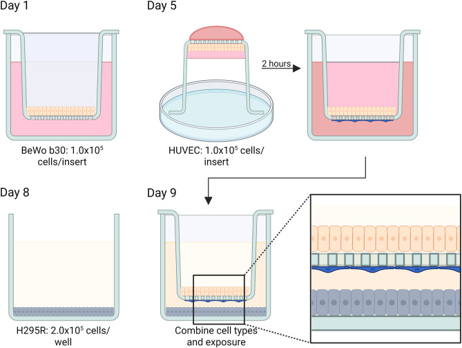
Their model mimics the structure of the barrier layers of a human placenta.
The first layer is made of BeWo b30 cells, which have similar qualities to the epithelial tissue that covers placental villi; the next layer is made from HUVEC cells, isolated from a human umbilical cord vein; and the third layer is of H295R cells, which originate from a human adrenal gland.
The BeWo b30/HUVEC cell layers were chosen to represent the physical placental barrier, while the H295R cell layer represents the main hormone-producing organ within the placenta, known as the fetal adrenal.
Once they’d set all this up, they started adding different kinds of micro- and nanoplastics to the ‘outer layer’, to see what would pass through the cells, and what impacts these particles would have on the hormones the cells produced.
Across 72 hours, most of the polymers had made their way right through all layers of the membrane; some more so than others. The only variety that was not detected in measurable amounts on the other side of the membrane was polyamide.
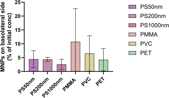
Fluorescence microscopy revealed there were more of these plastic particles embedded throughout the first two cell layers, though not the H295R cells.
Before even adding any plastics, the researchers detected 19 different steroid hormones in their tri-cell model. Despite some differing absolute hormone levels from pregnant humans in vivo, similar key steroidogenic pathways are active in the tri-culture model in this study.
Then, once the plastics were added, they could see which changes occurred.
Polymer exposure resulted in a shift in the cells that represent the outside of the model placenta. Multiple types of plastic resulted in a decrease in etiocholanolone, while polystyrene exposure affected estrogen levels.
Considering how important steroid hormones are in pregnancy, the researchers say further investigation is needed to understand the mechanisms underlying these results.
Related: Ovaries Appear to Develop an Incredible Second Role After Menopause
“Our study shows that several types of micro- and nanoplastics can cross a human placental model and influence hormone production,” van Boxel says.
“While this does not directly mean they cause harm during pregnancy, it highlights the urgent need to better understand how these particles may affect fetal development.”
The research was published in Molecular and Cellular Endocrinology.
This article was fact-checked by Rachel Garner and edited by Peter Dockrill. While we pride ourselves on our process, we are only human. If you spot a mistake, please let us know.