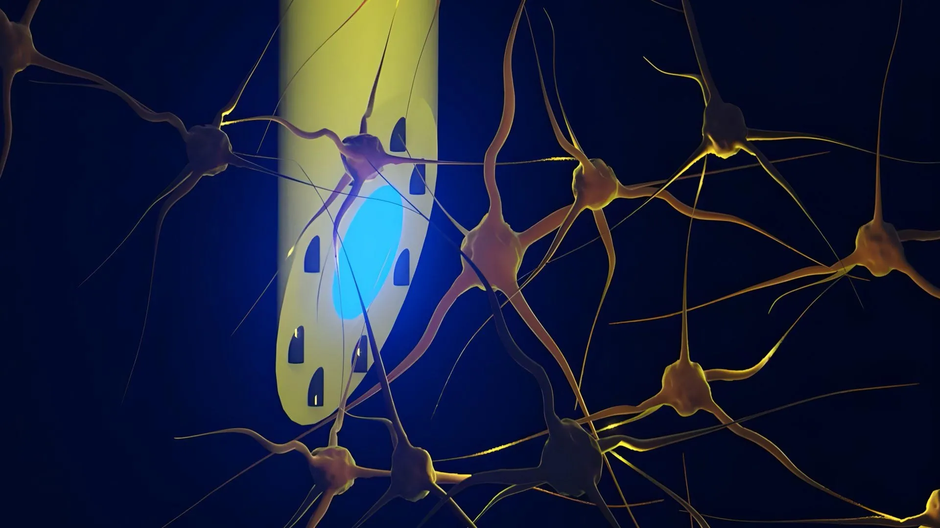
Эрдэмтэд тархины мэдрэлийн дохиог бүртгэх, эм тариа хүргэх, цахилгаан болон гэрлээр өдөөх үйлдлийг нэгэн зэрэг гүйцэтгэх чадвартай зүү мэт нарийн төхөөрөмжийг гарган авлаа.
DTU, Копенгагены их сургууль, Лондонгийн их сургуулийн коллежийн судлаачид “mAxialtrode” хэмээх микрофлюидик суулгацыг хөгжүүлж, үр дүнгээ Advanced Science сэтгүүлд нийтлүүлжээ. Энэхүү төхөөрөмж нь уян хатан, хуванцар шилэн утаснаас бүрдэх бөгөөд 0.5 мм-ээс бага диаметртэй тул тархины эдэд учруулах гэмтлийг багасгадаг. Уг суулгацын гол хэсгээр гэрэл дамжуулах төв байрлах ба түүнийг тойрсон найман бичил суваг нь шингэн эм дамжуулах болон цахилгаан идэвхжлийг хэмжих металл утас агуулдаг байна.
Амьд хулгана дээр хийсэн туршилтаар уг төхөөрөмж нь тархины өнгөц болон гүн хэсгүүдэд (тухайлбал, тархины бор гадар болон хиппокамп) мэдрэлийн эсийг гэрлээр өдөөх, мэдээлэл бүртгэх, түүнчлэн гурван миллиметрийн зайтай өөр өөр цэгүүдэд эм тариа хүргэх чадвартай болохыг тогтоожээ. Ингэснээр олон төхөөрөмж зэрэг суулгах шаардлагагүй болж, тархины үйл ажиллагааг илүү нарийвчлалтай судлах боломж бүрдэж байна.
Судлаач Кунян Суйгийн тайлбарласнаар, энэхүү технологи нь одоогоор судалгааны шатандаа яваа бөгөөд цаашид эпилепси зэрэг мэдрэлийн тогтолцооны өвчнийг эмчлэхэд ашиглах ирээдүйтэй юм. Гэсэн хэдий ч төхөөрөмжийг эмнэлзүйн практикт нэвтрүүлэхийн тулд нэмэлт туршилт, боловсруулалт хийж, зохих зөвшөөрлүүдийг авах шаардлагатай байна.
Дэлгэрэнгүйг эх сурвалжаас харах
↓Эх сурвалжийг нээх ↓
A new type of brain implant could give scientists a more precise way to study the brain and may eventually contribute to treatments for neurological conditions such as epilepsy.
Researchers from DTU, the University of Copenhagen, University College London, and other institutions have developed a long, needle-thin brain electrode equipped with microscopic channels. Known as the microfluidic Axialtrode (mAxialtrode), the device is designed to provide multiple functional points along the length of the implant. This allows researchers to record neural activity and deliver medication to specific locations across different parts of the brain.
The findings were published in the journal Advanced Science.
A Multifunction Tool for Brain Research
For now, the technology is primarily intended as a research tool. Scientists could use it to investigate how signals travel through different layers of the brain during processes involving epilepsy, memory, and decision-making.
Over the longer term, the researchers say the mAxialtrode could also have therapeutic applications. One possibility would be using the device to deliver drugs to precise locations while simultaneously applying electrical stimulation or light stimulation to selected areas of the brain.
Postdoc Kunyang Sui, who developed the mAxialtrode concept together with Associate Professor Christos Markos, says one of the main advantages is that several capabilities can be combined within a single implant. That could allow researchers to perform more precise experiments while reducing the need for multiple devices inserted into the brain.
“Most current brain implants are based on hard materials such as silicon, which can irritate the brain and trigger inflammatory reactions in the tissue. The new implant differs in that it is made of soft, plastic-like optical fibers and has a specially angled tip that makes it smaller and reduces the damage caused when it is placed in the brain,” says Kunyang Sui.
Sui cautions that the technology is still far from routine clinical use. Extensive testing, additional development, and regulatory approvals would be required before it could be used to treat patients.
Moving Beyond Conventional Optical Fibers
Brain researchers currently use flat-ended optical fibers in many experiments. These thin fibers, made from glass or plastic, can carry light into deep regions of the brain. They are frequently used in optogenetics, a technique in which specific nerve cells are activated using light.
Conventional fibers, however, have an important limitation. They typically interact with brain tissue only at the end of the fiber, meaning researchers can stimulate or monitor just one location at a time.
The outermost end is called the distal tip, or the “nose” of the fiber. Light is emitted and contact with brain tissue occurs only at this point. As a result, scientists may be limited to measuring or stimulating one brain layer at a time, even though many brain functions depend on communication among several layers and deeper structures.
How the New Brain Implant Works
The needle-thin mAxialtrode begins as a much larger polymer rod. Researchers heat the material and draw it into an extremely thin fiber, somewhat like producing a very fine strand of sugar, but with far greater precision.
A light-conducting core runs through the center of the fiber. Surrounding it are eight microscopic channels that can transport liquids. Those channels can also hold extremely thin metal wires used to measure electrical activity in the brain.
The finished fiber measures less than half a millimeter across. It is also highly flexible, allowing it to move along with brain tissue rather than pressing rigidly through it. This difference in stiffness could be important because harder implants can trigger inflammatory responses when they remain in the brain for long periods.
Tested in Living Mice
The researchers tested the system not only in the laboratory but also “in vivo,” meaning in living mice. The electrode was implanted in the animals’ brains and connected to light sources, recording equipment, and small pumps used to deliver fluids.
The experiments showed that the device could stimulate nerve cells using both blue and red light. At the same time, researchers were able to record electrical activity from shallow and deeper brain regions, including the cerebral cortex and hippocampus.
They were also able to inject different substances at separate depths, with delivery points spaced almost three millimeters apart. All of these measurements and forms of stimulation were carried out using a single lightweight fiber, which the mice were able to carry without any obvious signs of discomfort.
Potential Applications in Epilepsy and Neuroscience
The in vivo experiments and neurophysiological validation were performed in close collaboration with Associate Professor Rune W. Berg of the University of Copenhagen and Associate Professor Rob C. Wykes from University College London. Their contributions included expertise in analyzing neural circuits and models relevant to epilepsy.
The research team is now working to patent the technology behind the brain electrode. The scientists are also exploring what would be required to begin testing the device in patients within a clinical setting.