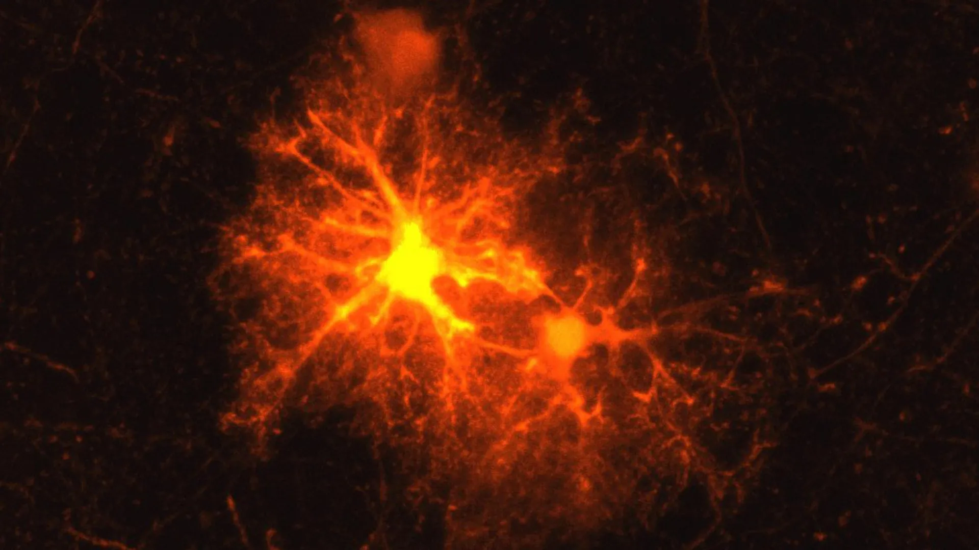
Олигодендроцит үүсгэгч эсүүд гэмтлийн дараа кортикотропин ялгаруулагч дааврыг идэвхжүүлж, тархины эд эсийн нөхөн төлжилтийг зохицуулдаг болохыг эрдэмтэд илрүүлжээ.
Max Planck Institute of Psychiatry-ийн судлаач Ян Дёссинг болон Клеменс Рис нар хулганы тархины гэмтлийн үед тодорхой бүлэг эсүүд идэвхжиж байгааг ажиглажээ. Шинжилгээгээр эдгээр эс нь миелин бүрхүүлийг үүсгэдэг олигодендроцит үүсгэгч эсүүд (OPCs) болохыг тогтоов. Миелин нь мэдрэлийн эсүүдийн хоорондох мэдээлэл дамжуулалтыг дэмжиж, тэжээлээр хангадаг тул тархины үйл ажиллагаанд чухал үүрэгтэй.
Судалгааны явцад гэмтлийн ойролцоох OPC эсүүдийн гуравны нэг нь стрессийн хариу урвалыг зохицуулдаг кортикотропин ялгаруулагч дааврыг (CRH) ялгаруулж эхэлдэг болохыг илрүүлжээ. Энэхүү үйл явц гэмтэл авснаас хойш хэдхэн цагийн дотор эхэлж, гурав орчим хоногийн дараа зогсдог нь CRH даавар эдгэрэлтийн эхний шатанд чухал үүрэгтэйг харуулж байна. Мөн CRH рецептор 1-ийн оролцоо нь OPC эсүүдийн боловсрох хугацааг хянаж, гэмтсэн миелинийг бүрэн нөхөн сэргээхэд зайлшгүй шаардлагатай гэж судлаачид үзэж байна.
Түүнчлэн эрдэмтэд уг систем тархины хөгжлийн явцад ч мөн адил үүрэг гүйцэтгэдэг болохыг тогтоожээ. CRH рецептор 1 дутагдалтай хулгануудын тархины бүтэц хөгжлийн явцад өөрчлөгдөж, миелин бүрхүүл илүү зузаарсан байна. Энэ нь CRH нь зөвхөн гэмтлийн дараах нөхөн сэргээлтэд бус, тархины анхдагч хөгжилд ч нөлөөлдөг байж болзошгүйг илтгэж байна.
Судлаачдын таамаглаж буйгаар, хөгжиж буй мэдрэлийн эсүүд CRH ялгаруулах замаар OPC эсүүдийн үржил болон олигодендроцит болон хувирах явцыг удирддаг аж. Цаашид энэхүү механизмыг судлах нь сэтгэл гутрал зэрэг стресстэй холбоотой сэтгэцийн эмгэгийг эмчлэх шинэ арга замыг нээж магадгүй юм. Судалгааны үр дүнг Cell Reports сэтгүүлд хэвлүүлжээ.
Дэлгэрэнгүйг эх сурвалжаас харах
↓Эх сурвалжийг нээх ↓
When laboratory mice experience brain damage, e.g., from an injection, Jan Deussing repeatedly notices the same response. A particular group of cells appears and becomes active around the damaged area. Although Deussing, a research group leader and experienced neurobiologist, had observed the phenomenon many times, he did not know exactly what type of cells were involved.
The mystery became an ideal research question for a master’s student. Clemens Ries, who had recently joined the Max Planck Institute of Psychiatry for an internship as he approached the end of his biology degree, took on the challenge.
Identifying the Brain’s Repair Cells
Using a mouse model, Ries systematically tested markers for all known cell types. Only one produced a response: the marker for oligodendrocyte progenitor cells (OPCs).
These precursor cells can mature into oligodendrocytes, which produce the myelin sheath surrounding axons. Axons are extensions of nerve cells that allow neurons to communicate with one another. Myelin acts much like the insulating material around an electrical cable. It supports efficient information transmission along axons and also helps supply them with nutrients, making it vital to healthy brain function.
Damage to myelin can have serious consequences. In autoimmune diseases such as multiple sclerosis (MS), the protective coating breaks down. Physical injuries can also harm myelin, and in severe cases, the resulting damage can lead to the death of entire neurons. Restoring myelin around affected axons is therefore an important part of the brain’s response to injury.
A Surprising Stress Hormone Appears After Injury
Ries initially studied the newly identified cells for his master’s thesis. “The topic remained so exciting that it became my doctoral thesis,” says the biologist.
His subsequent research showed that these precursor cells multiply dramatically around the edges of brain wounds. Most then continue to mature, eventually becoming oligodendrocytes capable of producing new myelin.
But Ries and Deussing also uncovered something that had not been known before. Near the damaged tissue, about one third of the OPCs activate corticotropin-releasing hormone (CRH), a hormone that plays a central role in regulating the body’s stress response. Researchers had not previously known that OPCs could produce neuropeptides such as CRH. The findings have now been published in the renowned journal Cell Reports.
The CRH response begins remarkably quickly. Production can be detected within just a few hours after an injury, but it shuts down again after roughly three days. This short and rapid burst suggests that CRH has an important function during the earliest stages of the healing response.
CRH Helps Control the Timing of Myelin Repair
One of the two known receptors for CRH also appears to be central to this process. CRH receptor 1 is present on a different population of OPCs and allows those cells to respond to the CRH that has been released.
When CRHR1 is absent, OPCs multiply more rapidly after an injury. That initial increase, however, does not translate into better repair. Ultimately, fewer mature oligodendrocytes are produced and remain.
The findings indicate that CRH helps regulate the timing of OPC maturation. That timing appears to be essential for producing enough mature oligodendrocytes to properly restore the damaged myelin sheath.
The Same System Shapes the Developing Brain
OPCs are not only important after injury. They also have a major role in building myelin as the brain matures. Much of this myelination takes place after birth and continues until young adulthood.
Because CRH receptor 1 is found on OPCs even when no injury is present, Ries and Deussing began to wonder whether the receptor might also influence myelination during normal brain development. Working with other researchers, they examined myelin formation in additional mouse models using several different methods.
They found that mice lacking CRH receptor 1 produced more OPCs during the early stages of development. Those changes did not disappear with age. Instead, they had lasting effects on the structure of the brain.
In adult brains, the researchers detected changes in myelination that could be traced to thicker myelin sheaths, particularly around thin axons. The results suggest that CRH receptor 1 on OPCs plays an important role not only in repairing myelin after injury, but also in regulating how myelin develops in the first place.
Where Does CRH Come From During Development?
Following an injury, OPCs themselves respond by producing and releasing CRH. Brain development raises a different question: where does the stress hormone come from when the brain is maturing normally?
The scientists propose that neurons may provide the answer. Their hypothesis is that developing neurons release CRH, which then influences both the multiplication of OPCs and their maturation into oligodendrocytes that produce myelin.
A Possible Connection to Depression and Stress
Neurons are already known to release CRH, particularly during stressful conditions. Stress experienced during early childhood development is also recognized as a risk factor for psychiatric disorders.
The new results therefore raise the possibility that the CRH system operating in OPCs could have broader implications for mental health.
“Our current findings suggest that in stress-associated psychiatric disorders such as depression, the CRH system in OPCs may play a greater role than previously known,” Deussing speculates.
If future research confirms and expands on that connection, understanding how CRH signaling affects OPCs, myelin formation, and brain development could eventually point toward completely new therapeutic approaches.